본문바로가기
닫기
About
Intro
Vision and Mission
Location
Academics
Undergraduate
Graduate
People
Faculty
Staffs
Research
Artificial Intelligence of Chemical Engineering
Energy & Environment
Polymers & Organic Materials
Biotechnology
Electronic Materials & Devices
News & Events
Research Highlight
Seminar
Notice
News
Support us & Donate
Alumni Association
Research Highlight
Home
News & Events
Research Highlight
Research Highlight
Seminar
Notice
News
Support us & Donate
Alumni Association
Research Highlight
총
58
건
제목
연세대 함승주 교수팀, 골드나노입자의 표면 개질과 효소 모방 작용을 동시 적용한 고민감 인플루엔자 A 바이러스 나노센서 개발 Professor Seungjoo Haam's team at Yonsei University has developed a highly sensitive influenza A virus nanosensor by simultaneously applying surface modification and enzyme-mimicking p
연세대학교(총장 윤동섭) 화공생명공학과 함승주 교수 연구팀이 골드나노입자의 표면 개질과 효소 모방 작용을 동시 적용한 고민감 인플루엔자 A 바이러스 나노센서에 대한 연구를 발표했다. 인플루엔자 A바이러스는 2019년부터 최근까지 전세계적 피해를 끼친 코로나 바이러스를 잇는 차세대 팬데믹의 유력 후보로 손꼽히고 있으며, 호흡기로 감염되어 빠르게 전파되는 만큼 조기 진단을 위한 고민감 현장형 센서의 중요성이 강조되고 있다. 이에 인플루엔자 A바이러스를 진단하는 다양한 연구들이 발표되고 있지만, 현장형을 띄는 비색 진단 도구로서 고민감도를 가지는 진단법에 대한 필요성이 여전히 강조되고 있다. 연세대 함승주 교수팀이 개발한 해당 진단 시스템은, 금 나노입자에 SH-PEG-biotin을 이용해 개질함으로써, biotin과 강하게 결합하는 단백질인 avidin을 골드입자와 순차적 처리를 진행해 타겟 바이러스 주위에서 layering을 형성하게 한다. 골드입자와 avidin의 순차적인 처리 cycle을 반복함으로써 적은 타겟 바이러스임에도 증폭된 신호를 얻을 수 있다. 더불어 골드나노입자가 가진 효소모방작용을 통해, 기질을 처리했을때, 골드입자가 가진 붉은색의 신호보다 더 증폭된 푸른색 신호를 얻을 수 있다. 따라서 이중으로 증폭된 신호를 얻음으로써 민감도가 높은 나노센서를 구현하였다. 연구팀이 개발한 해당 진단법은 3가지의 절차가 진행되는데 먼저 96 플레이트상에 항체를 코팅한 뒤, 타겟 바이러스가 sandwich 형태로 결합하는 pre-amplified step이다. 이때 타겟 바이러스에 결합하는 detection antibody에는 biotin이 결합 되어있다. 두번째로 avidin과 표면개질된 골드입자를 순차적으로 처리해주어 layering을 통한 신호 증폭을 이루는 1st amplification, 골드입자의 효소 모방작용으로 최종 신호 증폭을 이뤄내는 2nd amplification이다. 이러한 연쇄적인 증폭 효과로 101.29 EID50/mL 라는 우수한 LOD (limit of detection)을 얻을 수 있었으며, 이는 상용화된 rapid kit 보다 우수함을 보였다. 이번 연구는 과학기술정보통신부에서 주관한 나노 소재 기술개발사업과 환경산업부에서 주관한 실내공기 생물학적 위해인자 관리 기술개발사업의 지원을 받아 함승주 교수 연구팀의 정은지 연구원 (공동 제 1저자), 박근선 연구원(공동 제 1저자), 서울대 수의대 송대섭 교수 (공동 교신저자)와 함께 진행됐으며, 세계적인 과학 분야 권위지 (IF=15.9) ‘Small structures’에 24년 4월 7일(현지시간) 게재되었음과 더불어 연구의 우수성을 인정받아 표지 논문에 선정되었다. A research team led by Professor Seungjoo Haam from the Department of Chemical and Biomolecular Engineering at Yonsei University (President: Dongseop Yoon) has published a study on a highly sensitive influenza A virus nanosensor that applies surface modification of gold nanoparticles and enzyme-mimicking property. Influenza A virus is considered a candidate for the next global pandemic, following the SARS-CoV-2 virus that caused worldwide damage from 2019 to recent years. Given that it is a respiratory infection that spreads rapidly, the importance of high-sensitivity and on-site sensors for early diagnosis has been emphasized. Although various studies on diagnosing influenza A virus have been published, there is still a pressing need for highly sensitive colorimetric diagnostic tools suitable for field use. The diagnostic system developed by Professor Seungjoo Haam's team at Yonsei University involves modifying gold nanoparticles with SH-PEG-biotin, allowing for a strong bond with avidin, a protein with a high affinity for biotin. By sequentially treating the gold nanoparticles with avidin, they create a layering effect around the target virus. By repeating this cycle of sequential treatment with gold nanoparticles and avidin, the system amplifies the signal even when detecting a small amount of the target virus. Additionally, through the enzyme-mimicking property of gold nanoparticles, the system can produce a strong blue signal that is even more amplified than the red signal typically associated with gold nanoparticles when processing a substrate. This dual amplification approach results in a highly sensitive nanosensor with increased sensitivity to detect the influenza A virus. The system involves three key steps. First, they coat a 96-well plate with an antibody, creating a pre-amplification step where the target virus binds in a sandwich configuration. In this step, the detection antibody that binds to the target virus is conjugated with biotin. Next, in the 1st amplification, they achieve signal amplification by sequentially treating with avidin and surface-modified gold nanoparticles, resulting in a layering effect. Finally, in the 2nd amplification, the enzyme-mimicking action of the gold nanoparticles further amplifies the signal. This results in a final, more intense signal. This cascading amplification process leads to an impressive limit of detection (LOD) of 101.29 EID50/mL, demonstrating superior sensitivity compared to commercially available rapid test kits. The research was conducted by Eunji Jeong (co-first author), Geunsun Park (co-first author) of Professor Seungjoo Haam's research team, and Professor Daesup Song from Seoul National University's College of Veterinary Medicine (co-corresponding author) with the support of the the Nano Material Technology Development Program of the Ministry of Science and ICT and the Indoor Air Biological Hazard Management Technology Development Program of the Ministry of Environment. The research was published on April 7, 2024 (local time), in 'Small Structures,' a prestigious scientific journal with an impact factor of 15.9. Additionally, the study was selected as the cover article, further acknowledging its excellence. Small (2024) (IF: 15.9)Published: 07 April 2024https://doi.org/10.1002/sstr.202470014
연세대 함승주 교수팀, 바이러스와 융합이 가능한 플라즈모닉 나노 센서 개발 및 인플루엔자 A 바이러스 비색 진단에의 적용.Professor Seungjoo Haam's team at Yonsei University developed a plasmonic vesicle-mediated fusogenic immunoassay (PVFIA) for colorimetric detection of influenza A virus
연세대학교(총장 서승환) 화공생명공학과 함승주 교수 연구팀이 바이러스와 융합이 가능한 플라즈모닉 나노 센서를 개발하고 이를 인플루엔자 A 바이러스 진단에 적용하여 신속 정확한 면역분석법을 발표했다. 호흡기를 통해 전파되는 인플루엔자 바이러스는 빠른 전파 속도를 가질 뿐만 아니라 수많은 변종 출현 가능성으로 인해 전 세계적으로 심각한 피해를 일으키는 유해인자로 알려져 있다. 코로나 바이러스 출현 이후로 바이러스 진단 분야에서 민감하고 신속한 센서가 발표 되어 왔으며 이와 더불어 감염력이 있는 활성 및 온전한 바이러스 형태 (virion)을 인식할 수 있는 탐지 도구의 필요성이 강조되고 있다. 연세대 함승주 교수팀이 개발한 센싱 플랫폼은, 금 나노입자 (GNP)를 내부에 담지하고 있는 융합성 고분자 소포 (plasmonic vesicle, PV)를 개발하고 이와 더불어 표적 바이러스 특이적 포획을 위한 바이오칩을 적용함으로써, 바이러스와의 융합 및 선택적 결합 반응을 통해 비색 검출이 가능한 면역분석법 (PVFIA)이다. 연구팀이 개발한 PVFIA 분석법은 두 가지 순차적 분석이 수행되는데, 먼저 유리 기판에 항체를 코팅한 바이오 칩에 표적 바이러스의 선택적 포획을 선행하고 이어서 플라즈모닉 나노센서를 처리하여 포획된 표적 바이러스에 막융합을 유도하게 되면 내부의 금 나노입자가 유리 기판에서 색상 변화를 일으켜 시각적 검출이 가능하게 된다. PVFIA는 표적 IAV를 검출하는데 탁월한 특이성을 보이며, 융합 조건과 GNP는 상당한 색상 변화를 유도하기 때문에 두 가지 연속 분석을 통합하여 낮은 검출 한계 (100.8 EID50/mL)와 우수한 신뢰도 (0.99)로 기존 면역 분석보다 만 배 더 높은 감도를 확보했다. 나아가 PVFIA를 기반으로 한 비색 검출은 타액이나 비강액에 적용이 가능하여 추후 현장 진단에 유망한 도구로 이용될 수 있다. 이번 연구는 환경산업부에서 주관한 실내공기 생물학적 위해인자 관리 기술개발사업 및 과학기술정보통신부에서 주관하는 나노소재기술개발사업의 지원을 받아 함승주 교수 연구팀의 이소정 연구원(공동 제 1저자), 문예솔 연구원(공동 제 1저자), 서울대 수의대 송대섭 교수 (공동 교신저자)와 함께 진행됐으며, 세계적인 과학 분야 권위지 (IF=13.3) ‘Small’에 24년 1월 25일(현지시간) 게재되었음과 더불어 연구의 우수성을 인정받아 표지 논문에 선정되었다. A research team led by Prof. Seungjoo Haam of the Department of Chemical and Biological Engineering at Yonsei University (President Dong-seop Yoon) has developed a plasmonic vesicle-mediated fusogenic immunoassay (PVFIA) for colorimetric detection of influenza A virus. Influenza viruses, transmitted through the respiratory tract, are known to be a serious and damaging agent worldwide, not only their rapid spread but also because of the potential for numerous variants to emerge. Since the emergence of the coronavirus, sensitive and rapid sensors have been announced in the field of virus diagnostics, highlighting the need for detection tools that can recognize active and intact viral forms (virions) that are infectious. The sensing platform developed by the team of Professor Seungjoo Haam at Yonsei University is an immunoassay that enables colorimetric detection through fusion and selective binding reactions with viruses by developing fusible polymeric vesicles (plasmonic vesicles, PVs) containing gold nanoparticles (GNPs) and applying a biochip for targeted virus-specific capture. The PVFIA assay developed by the researchers involves two sequential assays: selective capture of target viruses on a biochip coated with antibodies on a glass substrate, followed by treatment of the plasmonic nanosensor to induce membrane fusion of the captured target viruses, where the internal gold nanoparticles cause a color change on the glass substrate, enabling visual detection. PVFIA shows excellent specificity in detecting target IAVs. Since the fusion conditions and GNPs induce significant color changes, integrating two consecutive assays resulted in a sensitivity ten thousand times higher than that of conventional immunoassays with a low detection limit (100.8 EID50/mL) and excellent reliability (0.99). Furthermore, colorimetric detection based on PVFIA can be applied to saliva or nasal fluids, making it a promising tool for point-of-care diagnostics in the future. This research was conducted by Dr. Sojeong Lee (first co-author) and Yesol Moon (first co-author) along with Prof. Seungjoo Haam’s research team, and Prof. Daesub Song (co-corresponding author) with the support of the Technology Development Project for Biological Hazards Management in Indoor Air Project funded by Korea Ministry of Environment and the Nanomaterial Technology Development Project promoted by the Ministry of Science and ICT. The work was published on January 25, 2024 (local time) in the prestigious journal of ‘Small’ and selected as a frontispiece cover. Small (2024) (IF: 13.3)Published: January 25, 2024https://doi.org/10.1002/smll.202305748
연세대 함승주 교수팀, 자가조립 서브유닛 백신 플랫폼 개발 자가조립 과정을 통해 효능을 개선한 고밀도 항원 분포 백신 입자 개발 성공 Professor Seung-Joo Haam’s team at Yonsei University developed a self-assembled subunit vaccine platform
연세대학교(총장 윤동섭) 화공생명공학과 함승주 교수 연구팀이 높은 면역원성을 가지는 자가조립 서브유닛 백신 플랫폼을 개발함으로써 전세계적으로 위협이 되고 있는 감염병에 대한 예방 대책 기술을 제시했다. 전염병은 공중 보건에 매우 심각한 영향을 끼칠 수 있으며, 실제로 2019년 말 발생한 COVID-19에 의해 그 심각성이 널리 알려진 바 있다. 따라서 미래에 잠재적으로 유행할 수 있는 전염병 (Disease X)을 예방하기 위한 백신 기술의 개발이 필요하다. 미래 백신 개발에 있어서는 백신 투여 과정에서 발생할 수 있는 다양한 부작용의 최소화와 효율적으로 면역 반응을 유도할 수 있는 높은 면역원성, 낮은 생산 비용 등이 주안점으로 꼽힌다. 연세대 함승주 교수팀이 개발한 자가조립 기반 서브유닛 백신 플랫폼은 양친매성 고분자와 접합된 항원으로 구성되어 있다. 양친매성 고분자는 수용성 용매와 유기 용매가 혼재하는 에멀젼 환경에서 자가조립 과정을 통해 입자형태로 구축되며, 유기 용매의 제거를 통해 표면에 항원이 고밀도로 존재하는 백신 입자가 제조된다.함승주 교수팀은 백신 플랫폼의 유효성을 다방면에서 검증하는 데에 성공했다. 면역 세포를 대상으로한 항원 전달능 및 수지상 세포의 활성화능의 개선을 확인하였으며, 동물실험에서 모델 항원 및 인플루엔자 바이러스 항원에 특이적인 항체의 생성량과 다양한 면역인자가 단일 항원 대비 증가하는 것 또한 확인하였다. 마지막으로, 인플루엔자 바이러스를 활용한 감염 실험에서 백신 투여 동물의 높은 생존률과 바이러스 농도 감소, T 세포 활성화를 확인하여 실제 바이러스의 예방에 활용될 수 있는 기술임을 보였다.특히, 이번 연구의 핵심은 자가조립 과정을 통한 표면 항원 밀도 증가 기술 개발에 있다. 기존 서브유닛 백신은 낮은 부작용과 생산 비용을 가지고 있으나, 면역원성 또한 낮은 한계점이 존재하여 이를 보완하기 위한 면역 보조제 (adjuvant)가 반드시 필요했다. 본 연구에서는 자가조립이 가능한 고분자를 활용하여 입자 표면에 고밀도의 항원이 분포하는 백신 입자를 개발하여, 별도의 면역 보조제 없이 서브유닛 백신의 낮은 면역원성을 극복했다. 연세대 함승주 교수는 본 연구를 통해 “COVID-19 판데믹 이후 감염병의 예방,치료,진단에 대한 수요가 커지고 있으며, 미지의 감염병 X에 빠르게 대응하기 위한 백신 플랫폼의 개발이 필요한 상황이다. 본 연구를 통해 개발된 백신 플랫폼은 항원의 교체가 용이하여 감염병 확산에 따른 조기 대응에 유리하며 서브유닛 백신의 단점을 극복했다. 또한, 본 연구성과를 통해 입자의 물리적인 특성을 통한 백신 기술 개발 분야에 적용 및 응용될 수 있을 것으로 기대된다”고 전했다. 이번 연구는 과학기술정보통신부가 지원하는 개인기초연구사업 및 신·변종감염병대응플랫폼핵심기술개발사업의 지원으로 함승주 교수 연구팀의 박근선 박사(공동 제1저자)에 의해 진행됐으며, 세계적인 과학 분야 권위지 ‘ACS Nano’에 1월 8일자(현지시간)로 게재 및 전면 표지로 선정되었다. Yonsei University’s (President Dong-seop Yoon) department of chemical and biomolecular engineering Professor Seung-Joo Haam’s research team developed a self-assembled subunit vaccine platform with high immunogenicity, providing a preventive technology against infectious diseases that pose a global threat. Infectious diseases can have a very serious impact on public health, which has been widely recognised by the COVID-19 outbreak in late 2019. Therefore, it is necessary to develop vaccine technologies to prevent potential future epidemics (Disease X). For future vaccine development, minimising various side effects that may occur during vaccine administration, high immunogenicity to efficiently induce an immune response, and low production costs are the main priorities. The self-assembly-based subunit vaccine platform developed by Seung-Joo Ham and colleagues at Yonsei University consists of antigens conjugated to amphiphilic polymers. The amphiphilic polymers are self-assembled into particles in an emulsion environment in which aqueous and organic solvents are mixed, and the removal of the organic solvent produces vaccine particles with a high density of antigen on the surface.Seung-Joo Ham's team has successfully validated the vaccine platform in a number of ways, including improved antigen delivery to immune cells and activation of dendritic cells, as well as increased production of antibodies specific to model antigens and influenza virus antigens in animal studies, as well as increased production of various immune factors compared to a single antigen. Finally, in infection experiments using influenza virus, we confirmed high survival rates, reduced viral concentrations, and T cell activation in vaccinated animals, indicating that the technology can be used to prevent actual viruses.In particular, the key to this research is the development of a technology to increase surface antigen density through a self-assembly process. Existing subunit vaccines have low side effects and low production costs, but they also have limitations in terms of immunogenicity, so an immune adjuvant is needed to compensate for this. This study developed vaccine particles with a high density of antigens distributed on the surface of the particles using self-assembling polymers to overcome the low immunogenicity of subunit vaccines without the need for an immune adjuvant. Professor Seung-Joo Haam of Yonsei University said, ‘After the COVID-19 pandemic, there is a growing demand for prevention, treatment, and diagnosis of infectious diseases, and it is necessary to develop a vaccine platform to quickly respond to unknown infectious diseases. The vaccine platform developed through this research overcomes the disadvantages of subunit vaccines by easily replacing antigens, which is advantageous for early response to the spread of infectious diseases and overcomes the shortcomings of subunit vaccines. In addition, it is expected that the results of this research can be applied to the field of vaccine technology development through the physical properties of particles.’ The research was conducted by Dr. Park Geun-seon (co-first author) of Professor Seung-joo Haam's research team with the support of the Individual Basic Research Project and the Core Technology Development Project for New and Variant Infectious Disease Response Platform supported by the Ministry of Science and ICT, and was published on 8 January (local time) in ACS Nano, a leading scientific journal, and was selected as the front cover. ACS Nano (2024) (IF: 17.1)Published: January 8, 2024https://doi.org/10.1021/acsnano.3c09672
연세대 함승주 교수팀, 자성체가 결합된 Polydiacetylene (PDA) 나노 입자를 이용한 마우스 소변내 HER2 과발현 유방암 유래 엑소좀의 비색검출. Professor Seung-Joo Haam's team at Yonsei University developed a magnetic-polydiacetylene nanoparticles for colorimetric detection of HER2-overexpressing-cancer-der
연세대학교 (총장 윤동섭) 화공생명공학과 함승주 교수 연구팀이 HER2 과발현 유방암 모델링 마우스 소변에서 비색변화를 통해 HER2 과발현 엑소좀을 선택적으로 검출하고 자석을 이용하여 성공적으로 분리했다. Polydiacetylene (PDA)은 다양한 외부 자극 (온도, pH, chemical 등등)에 의해 파랑색에서 붉은 색으로 변하는 특징을 가지는 양친매성 고분자이다. 양친매성 고분자는 좀 형태로 만들기 용이하기 때문에 PDA에 인지질을 추가하여 좀 형태의 나노 입자를 합성한 후 외부에 자성 나노입자 (magnetic nanoparticle)를 부착하여 자기장으로 분리가 가능하도록 센서를 설계하였다. 최종적으로, 합성된 자성 나노입자-PDA 복합체에 HER2 과발현 엑소좀과 특이적으로 반응할 수 있는 antibody를 개질하였다. 설계된 나노 센서는 HER2 과발현 엑소좀과 선택적으로 반응하여 푸른색에서 붉은색으로 비색 변화가 가능함과 동시에 외부에 결합된 자성 나노입자의 존재로 기존의 검출 센서가 검출된 엑소좀을 분리하지 못했던 한계를 극복하였다. 본 연구팀이 개발한 나노 센서는 타겟 엑소좀의 검출과 동시에 분리를 진행할 수 있는 새로운 종류의 센서이다. 마지막으로 유방암 모델링 마우스 소변을 이용하여 센서의 성능을 평가하였다. 본 연구팀이 개발한 자성 나노입자-PDA 복합체는 HER2 과발현 유방암 마우스의 소변에서만 붉은색으로 변하였고, 이것을 눈으로 확인할 수 있었다. 대조적으로 건강한 마우스 소변에서는 변화하지 않았습니다. 또한, 자석을 이용하여 HRE2 과발현 유방암 유래 엑소좀을 분리할 수 있었다. 특히, 이번 연구의 핵심은 기존 엑소좀 검출 센서들 처럼 타겟을 검출하는 것에서 끝나는 것이 아니라 분리를 동시에 할 수 있다는 것에 있다. 간단하고 직관적인 색 변화를 통한 검출로 의료 인프라가 열악한 저소득 및 중간 소득 국가 (LMIC; low-and middle-income countries )에서 값비싼 장비와 복잡한 절차를 대체할 수 있는 진단 플랫폼이 될 수 있을 것이라 기대된다. 또한, 이 플랫폼의 임상적 적용성을 명확하게 검증하기 위해 환자 샘플에 대한 성능 평가를 수행할 계획에 있다. 그리고 이 플랫폼에 다양한 항체를 적용하면 액체생검을 기반으로 한 다른 암이나 질병 진단에도 센서 확장이 가능하다. 연세대 함승주 교수는 “엑소좀의 검출과 동시에 분리를 진행하는 것은 질병 진단 및 치료과정에 있어 시간을 매우 단축할 수 있고, HER2 과발현 유방암의 현장 진단을 위한 액체생검 센서로서의 잠재력을 갖고 있으며 비전문가와 LMIC에서 쉽게 사용할 수 있을 것으로 기대된다”고 전했다. 이번 연구는 과학기술정보통신부가 추진하는 NRF를 통한 나노소재기술개발 사업의 지원으로 함승주 교수 연구팀의 김륜형 연구원과 함께 진행됐으며, 세계적인 과학 분야 권위지 ‘스몰 (Small)’에 표지로 23년 11월 14일자 (현지시간)로 게재됐다. Yonsei University’s (President Yoon, Dong Sup) department of chemical and biomolecular engineering Professor Seungjoo Haam's research team developed the a magnetic-polydiacetylene nanoparticles for colorimetric detection of HER2-overexpressing-cancer-derived exosomes in mouse urine. Polydiacetylene (PDA) is an amphiphilic polymer that changes color from blue to red in response to various external stimuli (temperature, pH, chemicals, etc.). Liposome-shaped nanoparticles were synthesized by adding lipids to PDA, an amphipathic polymer, and magnetic nanoparticles were attached to the outside to enable separation using a magnetic field. Finally, the synthesized magnetic nanoparticle-PDA complex was modified with an antibody that could specifically bind to exosomes overexpressing HER2. The designed nanosensor can selectively react with HER2-overexpressing exosomes and change colorimetrically from blue to red. Due to the presence of externally bound magnetic nanoparticles, the detected exosomes could be separated, thereby overcoming the limitations of existing exosome detection sensors. The nanosensor developed by our research team is a new type of sensor that can detect and simultaneously separate target exosomes. Finally, the performance of the sensor was evaluated using mouse urine modeling breast cancer. The magnetic nanoparticle-PDA complex developed by our research team turned red only in the urine of HER2-overexpressing breast cancer mice, and this could be confirmed with the naked eye. In contrast, there was no change in healthy mouse urine. Additionally, exosomes derived from HRE2-overexpressing breast cancer could be separated using a magnet. In particular, the key point of this research is that it can simultaneously perform target detection and separation, which existing exosome detection sensors were unable to do. We hope that detection through simple and intuitive color change can become a diagnostic platform that can replace expensive equipment and complicated procedures in low- and middle-income countries (LMIC) with poor medical infrastructure. Additionally, we plan to perform performance evaluations on patient samples to clearly validate the clinical applicability of this platform. Professor Seungjoo Haam of Yonsei University replied, “Detecting and simultaneously isolating exosomes can significantly shorten the time of disease diagnosis and treatment processes, has potential as a liquid biopsy sensor for point-of-care diagnosis of HER2-overexpressing breast cancer, and can be easily used even by non-specialists and in LMICs. And by applying various antibodies to this platform, the sensor can be expanded to diagnose other cancers or diseases based on liquid biopsy.” This research was conducted with Researcher Ryunhyung Kim (first author) of Professor Seungjoo Haam's research team with the support of the Nano-Material Technology Development Program through the NRF funded by the Ministry of Science and ICT. The work was published in cover the prestigious journal of ‘Small’ on 14 November 2023 (local time). Small (2024) (IF: 13.)Published: November 14 2023https://doi.org/10.1002/smll.202307262
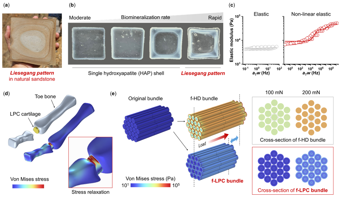
연세대 홍진기 교수팀, 바위에서 인공 관절의 해답을 찾다 Professor Jinkee Hong’s group at Yonsei University finds the solution to artificial joints from rocks
연세대학교(총장 윤동섭) 화공생명공학과 홍진기 교수 연구팀이 중앙대학교 기계공학과 이상민 교수 연구팀과 함께 인공 관절의 한계를 극복할 수 있는 소재를 개발했다. 관절염은 연골 손상으로 인한 점증적 또는 급성 발병으로 나타나 궁극적으로는 관절이 제 기능을 지 못하게 되는 질병이다.이를 치료하기 위해 물리치료, 운동요법 등 보존적 치료를 우선적으로 실시하나 증상이 호전되지 않을 경우, 수술적 치료로서 인공 관절 삽입이 시행된다. 기존의 인공 관절 소재는 세라믹, 폴리에틸렌 등의 견고한 물질을 사용하여 내구성이 뛰어나지만, 마찰계수가 높아 반복적인 움직임 시 마찰 부위에 피로가 누적되어 인공 관절의 손상이 일어나는 문제점이 있었다. 이를 해결 하기 위해 하이드로겔 등 유연한 소재를 활용할 수 있으나, 인체의 체중을 견디기에는 기계적 물성이 낮다는 문제점이 존재한다. 연세대 홍진기 교수팀은 이와 같은 문제를 해결하기 위해 바위에서 발견되는 나이테와 유사한 구조의 리제강 패턴(Liesegang Pattern)에 주목했다. 연구팀은 뼈의 주성분인 수산화인석이 하이드로겔에서 리제강 패턴을 지닌 형태로 생성되도록 하였으며, 이를 통해 하이드로겔의 유연한 특성을 유지하면서도 하중 분산 능력이 높은 비선형 탄성 (Nonlinear Elasticity)을 가진 하이드로겔을 개발했다. 인체의 움직임이나 충격을 완충하고 지지하는 데 도움이 되는 비선형 탄성은 인체 연골의 독특한 특성으로, 연골과 기계적 특징이 굉장히 유사한 이 개발 신소재는 인공 연골 모사 환경에서 놀라운 내구성을 보여줬다. 홍진기 교수는 “흔히 접할 수 있는 바위에서 착안해 의료 분야의 치명적인 문제에 해답이 되는 기술을 개발한 이상적인 화학공학 연구 결과물”이라고 평가하며, “특히 손가락의 인장 관절, 목의 추축 관절, 손목의 타원형 관절 등에 이 신소재가 유용할 것을 기대하며, 이를 향한 전임상 연구를 기획 중이다.”라고 덧붙였다. 본 연구는 한국연구재단(NRF), 과학기술정보통신부(MSIT), 국가신약개발사업단, 산업통상자원부(MOTIE)의 지원을 받아 진행됐으며, 연구 결과는 재료 분야 국제 최고 권위 학술지 ‘사이언스 어드벤시스(Science Advances)’에 4월 26일 게재됐다. Yonsei University’s (President Dong-Sup Yoon) department of chemical and biomolecular engineering Professor Jinkee Hong's research team developed a material that can overcome the limitations of artificial joints. Arthritis is a disease characterized by progressive or acute onset due to cartilage damage, ultimately leading to the joint's inability to function properly. To treat this condition, conservative treatments such as physical therapy and exercise therapy are prioritized. However, if symptoms do not improve, surgical treatment involving the insertion of artificial joints is performed. Conventional artificial joint materials, such as ceramics and polyethylene, are durable but suffer from the problem of cumulative fatigue at frictional sites due to their high friction coefficients during repetitive movements, leading to artificial joint damage. To address this issue, flexible materials such as hydrogels can be used, but they have low mechanical properties to withstand the body's weight. Professor Jinkee Hong’s team at Yonsei University addressed this problem by focusing on Liesegang patterns, similar to those found in rocks. The research team enabled hydroxyapatite, a major component of bones, to form Liesegang patterns within hydrogels, resulting in the development of hydrogels with high load distribution capabilities and nonlinear elasticity while maintaining the flexible properties of hydrogels. Nonlinear elasticity, which helps cushion and support the body’s movements and impacts, is a unique characteristic of human cartilage. This newly developed material, which mimics the mechanical characteristics of cartilage, showed remarkable durability in artificial cartilage simulation environments. Professor Jinkee Hong said “This is an ideal result of chemical engineering research which developed a strategy to solve the critical problems in the medical field by taking inspiration from Nature’s wisdom.” He also said, “In particular, we expect this new material to be useful for the tensile joints of the fingers, the pivot joint of the neck, and the elliptical joint of the wrist, and we are planning preclinical research toward this”. This research was conducted with support from the National Research Foundation of Korea (NRF), the Ministry of Science and ICT (MSIT), the National New Drug Development Project, and the Ministry of Trade, Industry and Energy (MOTIE), and the paper was published in Science Advances on April 26. Science Advances (2024) (IF: 13.6)Published: April 26, 2024https://www.science.org/doi/10.1126/sciadv.adl3075
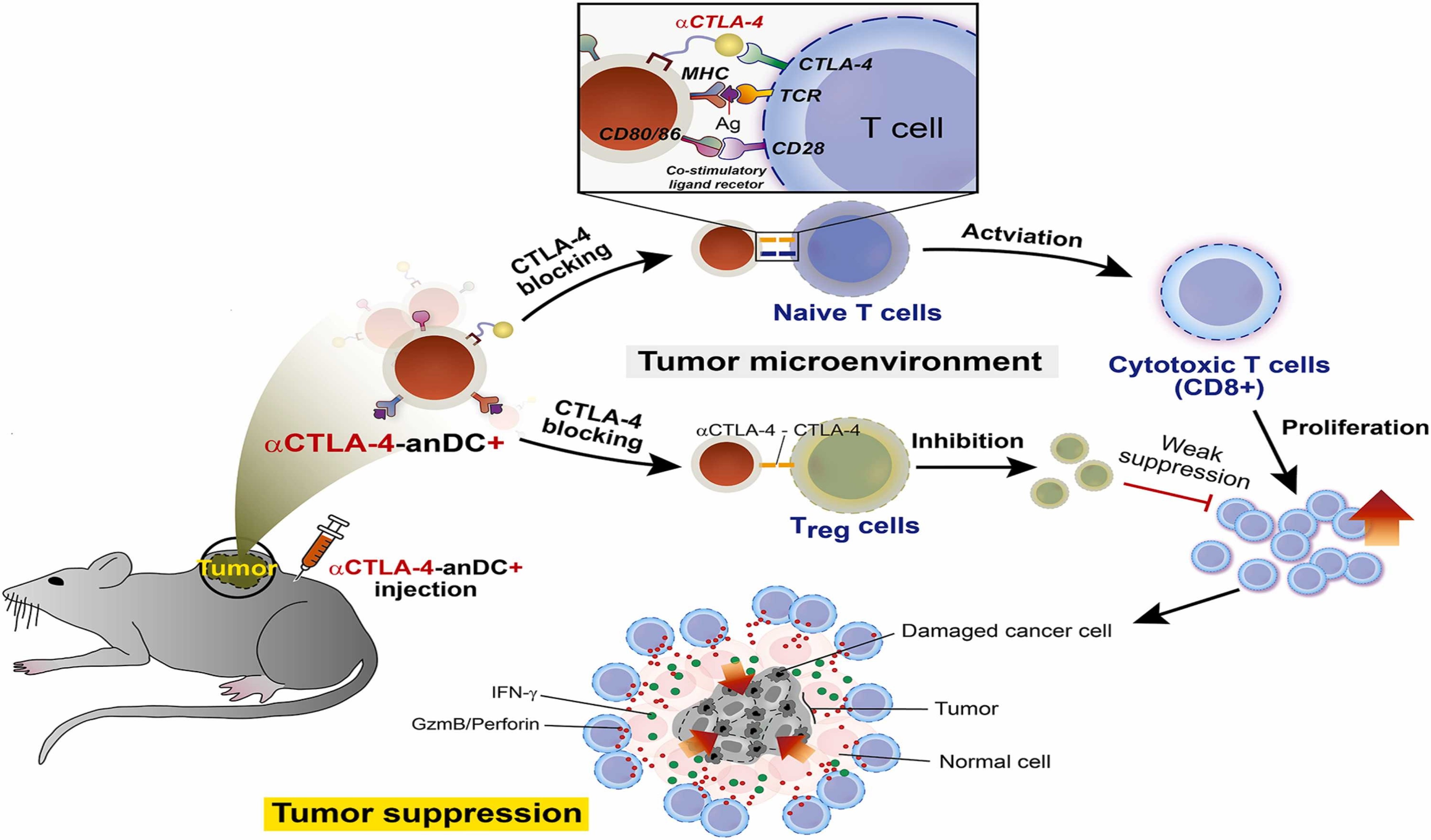
연세대 홍진기 교수팀, 인공 나노 수지상세포를 이용한 차세대 암 백신 플랫폼 개발 Professor Jinkee Hong’s group at Yonsei University developed a Next-Generation Cancer Vaccine Platform Using artificial nano Dendritic Cells.
연세대학교(총장 윤동섭) 화공생명공학과 홍진기 교수 연구팀이 전통적인 수지상세포 기반 암면역 치료의 한계 극복을 위해 ‘인공 나노 수지상세포(aritificial nano Dendritic Cell; 이하 anDC)’를 개발하였다. 개발된 암 백신 플랫폼은 다양한 암항원 탑재가 가능하며 대량생산 및 지속보관이 가능하기 때문에 다양한 암종에 대한 면역치료법 개발에 활용될 수 있다. 수지상세포(dendritic cell, DC)는 선천면역 및 후천면역을 모두 유도할 수 있는 면역계의 가장 핵심적인 항원제시세포(antigen-presenting cell, APC)로써 T세포 활성화에 필수적이다. 자가유래 DC를 이용한 DC 암 백신인 Provenge는 2010년 전이성 전립선암 치료제로 미국 FDA에서 승인되었으나, 짧은 체내 반감기, 낮은 보조 자극 능력, 그리고 높은 투여량과 잦은 투여 횟수에 따른 비용 증가의 한계로 현재는 임상에서 사용되지 않고 있다. PD-1 항체를 비롯한 면역관문 억제제는 약 20종의 암종에 대해 11종의 면역관문 억제제가 승인 되었고 이를 포함한 면역 항암 치료는 임상에서 활발히 사용되고 있다. 하지만 고형암에 대한 PD-1 저해제는 암종 및 환자에 따른 효능 차가 커서 불용성을 극복하기 위한 새로운 전략 도출이 필요하다. 연세대 홍진기 연구팀은 기존 DC 암 백신과 면역관문 억제제의 한계를 극복하기 위해 나노기술을 접목하여 새로운 형태의 anDC 암 백신을 개발하였다. DC의 T세포 활성화에 필수적인 다양한 분자가 세포막에 존재한다는 사실에 착안하여 DC 세포막을 금나노입자에 부착시킴으로써 대량생산 및 장기 보관이 가능한 anDC 암 백신 플랫폼을 완성하였다. 또한 제작된 anDC 암 백신에 CTLA-4 항체를 접합시킴으로써 T세포 활성 최적화를 유도하고자 하였다. CTLA-4 항체 접합 anDC는 마우스에 투여되었을 때 매우 효과적으로 암 생성을 저해하였고 한달 이상의 장기 보관 후에도 효능이 지속되었다. 이러한 강력한 항암면역반응은 anDC 투여로 암세포를 살상하는 T세포 빈도의 증가와 T세포 면역반응을 억제하는 조절T세포의 감소에 의한 것임을 확인하였다. 홍진기 교수는 “본 연구팀이 개발한 새로운 플랫폼의 암 백신은 기존 DC 암 백신의 한계를 극복할 수 있을 뿐만 아니라 암항원 특이적 T세포를 표적화할 수 있어 최근 임상시험 중인 mRNA 암 백신의 단점도 동시에 극복할 수 있다”라고 본 기술의 강점을 제시했다. 또한, “나노기술의 접목으로 자가유래 anDC 뿐만 아니라 Off-the-Shelf 형태의 anDC 제작이 가능하기 때문에 개인 맞춤형이자 기성품 형태의 항암제로의 개발이 가능하다”라고 전망하였다. 본 연구는 국가신약개발재단 국가신약개발사업과 한국연구재단 국제협력사업의 지원으로 수행되었다. 이번 연구는 홍진기 교수 연구팀의 최다희 박사(제1저자), 김태현 박사과정생(제1저자)이 연세대학교 생화학과 하상준 교수(공동 교신저자) 연구팀의 강태건 박사(제1저자), 문채원 박사과정생(제1저자)과 함께 진행하였으며, 세계적인 과학 분야 권위지 ‘나노 투데이 (Nano Today, IF 17.4)’에 3월 27일자(현지시간)로 온라인 게재됐다. 또한 본 연구의 핵심 기술에 대한 특허는 ㈜포투가바이오 사로 기술이전되어 면역항암치료제 플랫폼에 대한 후속 개발이 진행되고 있다. The research team led by Professor Jinkee Hong at Yonsei University's Department of Chemical and Biomolecular Engineering (President Dong-Sup Yoon) has developed an 'artificial nano Dendritic Cell (anDC)' to overcome the limitations of traditional dendritic cell-based cancer immunotherapy. The developed cancer vaccine platform can carry various cancer antigens and is capable of mass production and long-term storage, making it applicable for the development of immunotherapies for various types of cancer. Dendritic cells (DC) are essential antigen-presenting cells (APC) in the immune system, capable of inducing both innate and adaptive immunity, and are crucial for T cell activation. Provenge, a DC-based cancer vaccine using autologous DCs, was approved by the U.S. FDA in 2010 for the treatment of metastatic prostate cancer. However, it is currently not in clinical use due to its short in vivo half-life, low co-stimulatory capability, and the increased cost associated with high dosages and frequent administrations. Immunotherapy, including immune checkpoint inhibitors such as PD-1 antibodies, has been approved for about 20 types of cancer with 11 kinds of immune checkpoint inhibitors and is actively used in clinics. However, PD-1 inhibitors for solid tumors require new strategies due to significant efficacy differences between cancer types and patients. To overcome the limitations of existing DC cancer vaccines and immune checkpoint inhibitors, Yonsei University's Jinkee Hong research team has developed a new type of anDC cancer vaccine by incorporating nanotechnology. Based on the presence of various molecules essential for T cell activation on the DC membrane, the team attached the DC membrane to gold nanoparticles to create a mass-producible and long-term storable anDC cancer vaccine platform. Furthermore, they aimed to optimize T cell activation by conjugating the CTLA-4 antibody to the manufactured anDC cancer vaccine. The CTLA-4 conjugated anDC proved highly effective in inhibiting cancer formation when administered to mice and maintained its efficacy after long-term storage for over a month. This strong anti-cancer immune response was confirmed to result from increased T cell frequency killing cancer cells and decreased regulatory T cells suppressing the immune response. Professor Jinkee Hong stated, "Our newly developed platform not only overcomes the limitations of existing DC cancer vaccines but also targets cancer antigen-specific T cells, simultaneously overcoming the disadvantages of recently clinically tested mRNA cancer vaccines." He also predicted, "With the integration of nanotechnology, not only autologous anDCs but also Off-the-Shelf anDC production is feasible, allowing for the development of personalized yet ready-made cancer treatments." This research was supported by Korea Drug Development Fund and the framework of international cooperation program managed by the National Research Foundation of Korea. The research was conducted by Dr. Daheui Choi (first author) and PhD student Taihyun Kim (first author) from the research team of Professor Jinkee Hong, together with Dr. Tae-Gun Kang (first author) and PhD student Chae-Won Moon (first author) from the research team of Professor Sang-Jun Ha (corresponding author) in the Department of Biochemistry at Yonsei University, and was published online in the prestigious scientific journal 'Nano Today (IF 17.4)' on March 27th. Moreover, the core technology of this research has been transferred to Fortugabio Inc. for further development of the immunotherapy platform. Nano Today (2024) (IF: 17.4)Published: March 27, 2024https://www.sciencedirect.com/science/article/pii/S1748013224000926
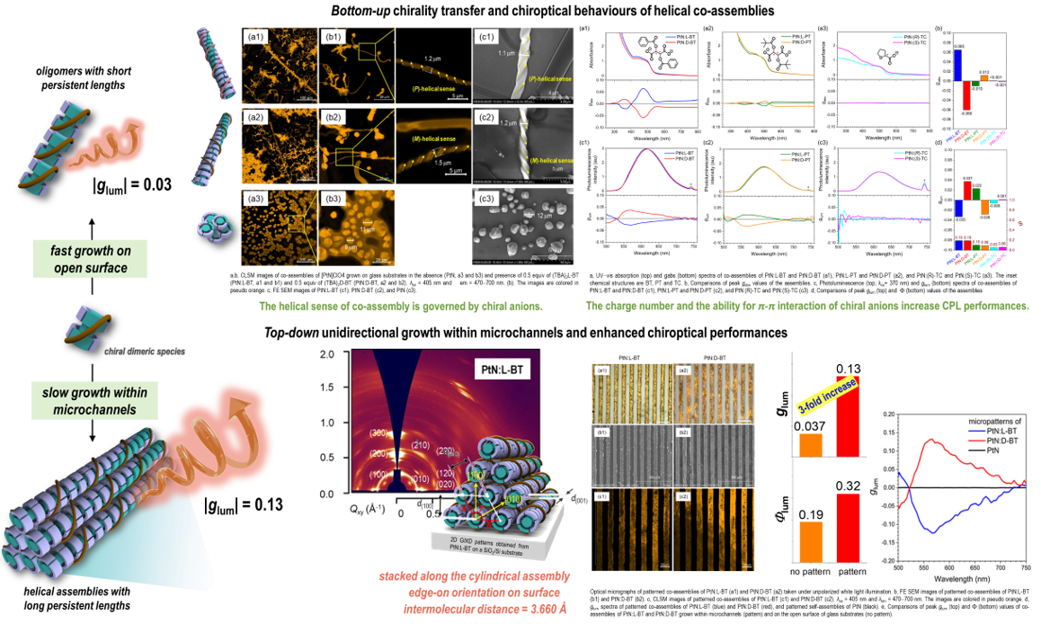
연세대 유영민 교수팀, 원편광 발광 효율을 극대화할 수 있는 신규 초분자 시스템 개발 Professor Youngmin You’s group at Yonsei University developed a novel approach to the highly efficient circularly polarized phosphorescence enhancement
연세대학교(총장 윤동섭) 화공생명공학과 유영민 교수 연구팀이 원편광 발광 특성을 극대화할 수 있는 신규 재료 시스템을 개발했다. 원편광은 오른쪽 또는 왼쪽으로 회전하는 편광축을 가지고 있는 빛을 의미한다. 원편광을 자체적으로 발광하는 재료나 시스템은 3D 디스플레이, 암호화 소자, 양자 통신, 비대칭 광합성, 스핀트로닉스 등 미래 혁신 기능성을 가지는 응용 가능성을 가지고 있어 최근 많은 연구가 이루어지고 있으나, 기본적으로 발광 효율과 원편광 발생 비율 사이 존재하는 트레이드 오프 관계 때문에 고성능의 원편광 발광 시스템을 구현하는 것이 어렵다. 연세대 유영민 교수팀은 인광을 발광하는 양이온 백금 착체와 키랄성 음이온 재료를 개발하였다. 평면 사각 분자 구조를 가지는 백금 착체는 금속 d 오비탈 겹침을 통한 초분자체 형성에 유리하고, 키랄성 음이온 재료를 섞어줌으로써 키랄 구조에 따라 나선 방향이 조절된 초분자체가 형성됨을 규명하였다. 개발한 신규 나선형 백금 초분자체 형성 전략은 기존의 원편광 발광 시스템이 가지고 있던 트레이드 오프 문제를 극복하여 백금 착체 단분자 대비 발광 효율과 원편광 발생 비율이 동시에 증진되는 특성을 보였으며, 마이크로 채널 안에서 초분자체를 형성하여 초분자체 간의 상호작용을 극대화함으로써 매우 높은 정렬도를 가지는 어레이를 soft-lithography 방식을 통해 구현했다. 이를 통해, 기존 보고된 원편광 발광 시스템 중에서도 구현이 힘들었던 30% 이상의 발광효율과 0.13의 원편광 발광 비대칭 인자 수치를 동시에 가지는 고효율 원편광 발광 시스템을 제안하였다. 이 연구를 통해 원편광 발광 극대화의 신규 분자 디자인 전략 및 미래 지향형 디스플레이 재료 개발이 가능할 것으로 예상된다. 본 연구성과는 한국연구재단 (NRF) 및 삼성 미래기술 육성센터의 지원을 받아 수행되었다. 이번 연구는 유영민 교수 연구팀의 박규림 석사 (제 1저자)가 함께 진행하였으며, 국제 저명 화학학술지인 Angewandte Chemie, International Edition (Impact Factor: 16.6)에 “Enhancing Circularly Polarized Phosphorescence via Integrated Top-Down and Bottom-Up Approach” 라는 제목으로 온라인 게재됐다. A research team led by Professor Youngmin You from the Department of Chemical and Biomolecular Engineering at Yonsei University (President Yun Dong-seop) has developed a new material system that maximizes the properties of circularly polarized luminescence (CPL). Circular polarized light refers to light whose polarization axis rotates either to the right or the left. Materials or systems that emit circularly polarized light inherently have potential applications in 3D displays, cryptographic devices, quantum communications, asymmetric photosynthesis, and spintronics, leading to significant research interest. However, implementing high-performance circularly polarized luminescence systems has been challenging due to the inherent trade-off between luminescence efficiency and the circular polarization ratio. Professor Youngmin You’s team developed a system using cationic Pt(II) complex that emit phosphorescence and chiral anionic materials. The square planar Pt(II) complex structure facilitates the formation of supramolecular structures through the overlap of metal d-orbital. By mixing with chiral anionic materials, the helical sense of supramolecules can be successfully controlled, yielding CPL. This novel strategy forming helical Pt(II) assembly overcame the trade-off problem by enhancing both photoluminescence efficiency and dissymmetry factor of CPL, which surpass the previously reported CPL system. By constructing helical Pt(II) assemblies within microchannels by soft-lithography method afford tight and well-aligned crystalline helical assemblies. This resulted in a high-efficiency circularly polarized luminescence system that features over 30% photoluminescence quantum efficiency and a dissymmetry factor of CPL of 0.13, which are exceptional figures among previously reported systems. This research suggests new molecular design strategies for maximizing circularly polarized luminescence and potential development of future-oriented display materials. The study was supported by the National Research Foundation of Korea (NRF). It was conducted by Gyurim Park (first author) of the team. The research has been published online in the prestigious journal Angewandte Chemie, International Edition, under the title "Enhancing Circularly Polarized Phosphorescence via Integrated Top-Down and Bottom-Up Approach." Angewandte Chemie, International Edition (2024) (IF: 16.6)Published: October 03, 2023https://doi.org/10.1002/anie.202309762
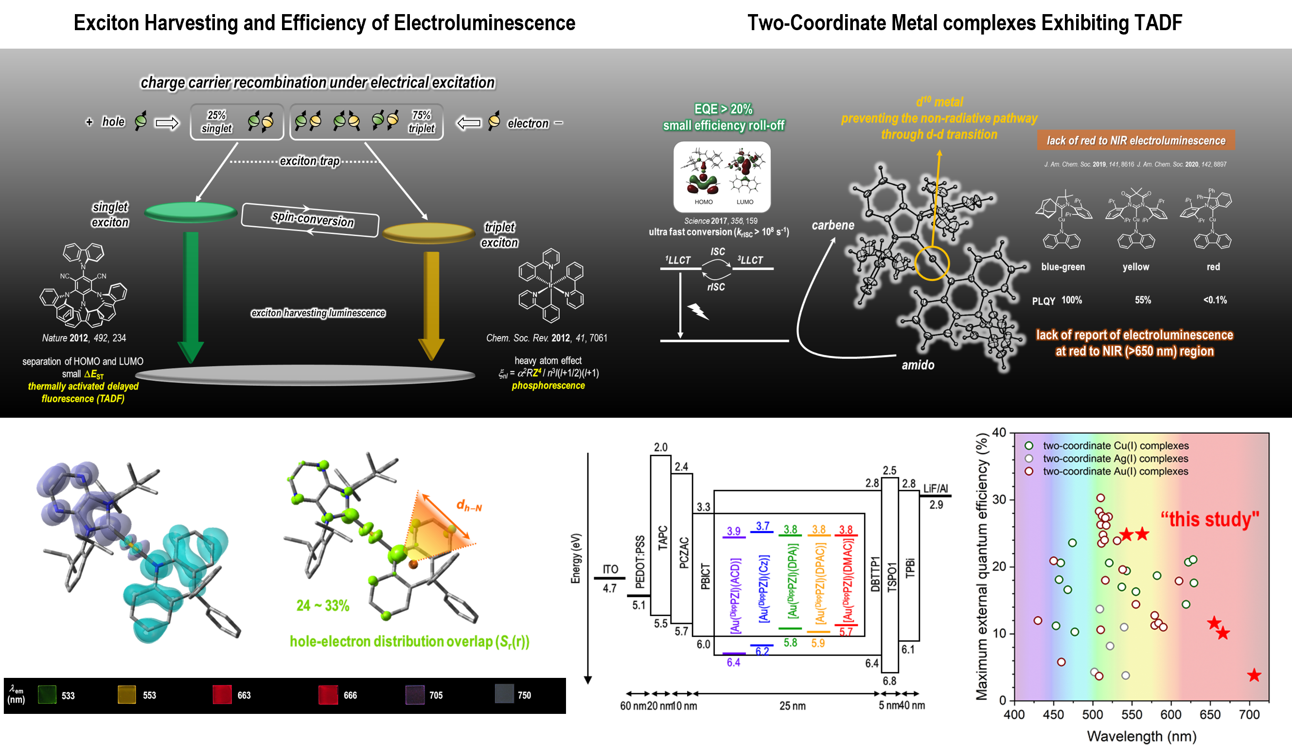
연세대 유영민 교수팀, 에너지 갭 법칙을 극복하는 신규 고효율 적색-근적외선 전계발광 2배위 금 착체 개발 Professor Youngmin You’s group at Yonsei University developed a highly efficient electroluminescent two-coordinate Au(I) complexes overcoming energy gap law
연세대학교(총장 윤동섭) 화공생명공학과 유영민 교수팀이 고효율의 적색-근적외선 영역 유기 발광 다이오드 (Organic Light Emitting Diodes (OLEDs)) 에 사용될 수 있는 신규 2배위 금 착체 분자를 개발하였다. OLEDs에 사용되기 위한 기존 유기 발광재료의 조건으로는 주입되는 전자쌍을 100% 빛으로 전환하는 능력이 필요하고 이를 위해 여기상태에서의 전자 스핀 변환이 가능한 발광 재료가 필요하다. 기존의 재료들은 이리듐이나 백금 등의 희토류 금속을 사용하는 인광 발광 소재 혹은 순수 유기물로 구성된 열활성 지연형광 재료들이 주로 연구되어 왔다. 그러나, 희토류 금속 기반의 발광재료는 가격적인 측면과 재료 매장량에 따른 한계가 분명하고 순수 유기물 기반 열활성 지연형광 재료는 느린 전계 발광 거동으로 소자 수명 측면에서 명확한 단점을 나타내고 있다. 최근에는 이와 같은 문제를 해결하고자 주화 금속 (금, 은, 동) 기반의 신규 2배위 유기금속착체들이 고효율 OLEDs 발광 소재로 부상하고 있다. 해당 소재들은 전자 주개와 전자 받개의 역할을 하는 리간드를 포함하여 리간드 간 전자 전이를 통해 열활성 지연 형광 특성을 나타내면서도 중심 금속에 원자 크기가 큰 금을 도입하여 빠른 전자 스핀 변환 속도를 가져, OLEDs 수명 문제를 해결할 유력한 후보 물질들로 여겨지고 있다. 연세대 유영민 교수팀에서는 리간드 구조의 조절을 통해 적색과 근적외선 발광 영역의 신규 2배위 금 착체들을 개발하였다. 기존의 OLEDs 발광 재료의 경우 장파장 영역에서 발광 효율이 급격히 감소하는 밴드갭 에너지 법칙을 따르고 있어 높은 발광 효율을 구현하기 어려운 문제가 있다. 유영민 교수팀에서는 개발한 신규 발광 재료들의 구조적 특성, 광물리 거동 및 양자화학계산에 기반한 분석을 통해 밴드갭 에너지 법칙을 극복할 수 있는 새로운 분자 디자인 원리를 제시하였다. 최종적으로, 개발된 발광 재료는 성균관대학교 이준엽 교수팀과의 공동연구를 통해 OLEDs 소자에 적용이 되었고, 성공적으로 적색에서 근적외선 영역에 이르는 발광 특성과 근적외선 영역에서도 외부 양자 효율 10%에 근접하는 높은 소자 효율을 달성하였다. 특히, 유영민 교수 연구팀에서는 해당 연구는 기존의 2배위 주화 금속 착체에서는 보고되지 않았던 650 nm 이상의 장파장 영역의 OLEDs 소자 특성을 새로 보고함으로써 기존 분자 특성의 한계를 넘을 수 있는 새로운 분자 디자인의 단서를 제공하는데 큰 의의가 있을 것으로 연구 의의를 전했다. 본 연구성과는 한국연구재단 (NRF) 의 지원을 받아 수행되었다. 이번 연구는 유영민 교수 연구팀의 Sreenivas Avula 박사 (제 1저자) 와 전병학 박사 (제 1저자)가 함께 진행하였으며, 국제학술지 어드밴스드 사이언스 (Advanced Science, Impact Factor: 15.1)에 “Achieving Long-Wavelength Electroluminescence Using Two-Coordinate Gold(I) Complexes: Overcoming the Energy Gap Law” 라는 제목으로 온라인 게재됐다. Professor Youngmin You and his team at the Department of Chemical and Biomolecular Engineering, Yonsei University (President Yoon Dong-sup), have developed a novel two-coordinate gold(I) complexes for highly efficient organic light emitting diodes (OLEDs) showing red-to-near-infrared (NIR) electroluminescence. For highly efficient OLEDs, the luminescent materials should convert 100% of injected electron pairs into light. This is facilitated by enabling electron spin conversion in the excited state, essential for achieving 100% of internal quantum efficiency. Up to now, phosphorescent transition metal complexes (i.e., Ir, Pt, and etc.) and purely organic materials showing thermally activated delayed fluorescence (TADF) have been considered as highly efficient electroluminescent materials. However, rare earth metals like iridium are constrained by high costs and supply issues, while organic TADF materials face challenges in device longevity due to their slow spin conversion rates. Recently, two-coordinate metal complexes based on coinage metals (i.e., Cu, Ag, and Au) have emerged as novel candidate for highly efficient OLED luminescent materials. These materials, which include ligands acting as electron donors and acceptors, display TADF through ligand-to-ligand charge transfer and having ultrafast spin-conversion rate originated from high spin-orbit coupling by metal center, making them strong candidates for resolving OLED lifespan issues. Professor Youngmin You’s team at Yonsei University has developed novel two-coordinate gold complexes for red to NIR luminescence. Current OLED luminescent materials often struggle to achieve high electroluminescence efficiency in the long-wavelength region, as they typically exhibit a significant decline in photoluminescence efficiency governed by the bandgap energy law. The research team has proposed a new molecular design principle that overcomes the bandgap energy law based on the structural characteristics, photophysical properties, and quantum chemical calculations of the newly developed luminescent materials. Finally, the series of gold complexes were applied to OLED devices in collaboration with Professor Jun Yeob Lee’s team at Sungkyunkwan University, achieving highly efficient electroluminescence having an external quantum efficiency of nearly 10% in the near-infrared region. In particular, Professor Youngmin You’s research team reported, for the first time, OLED devices in the long-wavelength region beyond 650 nm, a milestone previously unachieved with two-coordinated metal complexes. This research was supported by the National Research Foundation (NRF) of Korea. The research was conducted by Dr. Sreenivas Avula and Dr. Byung Hak Jhun of Yonsei University as the first authors. This research was published online in the international academic journal Advanced Science (Impact Factor: 15.1) under the title "Achieving Long-Wavelength Electroluminescence Using Two-Coordinate Gold(I) Complexes: Overcoming the Energy Gap Law". Advanced Science (2024) (IF: 15.1)Published: January 05, 2024https://doi.org/10.1002/advs.202305745
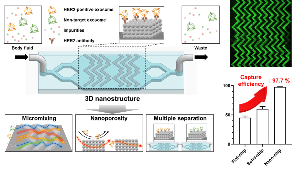
연세대 함승주 교수팀, 헤링본 패턴으로 배열된 나노구조체 기반 엑소좀 포집 미세유체 칩 개발. Professor Seungjoo Haam's team at Yonsei University developed an exosome capture microfluidic chip based on nanostructures arranged in a herringbone pattern.
연세대학교(총장 윤동섭) 화공생명공학과 함승주 교수 연구팀이 HER2 양성 유방암 유래 엑소좀을 포집할 수 있는 헤링본 패턴으로 배열된 3D 나노구조를 가지는 미세유체 칩을 개발했다. 이는 다양한 체액(혈장, 소변 등) 내에 존재하는 엑소좀을 높은 효율로 분리함으로써, HER2 과발현 유방암을 진단하고 모니터링 할 수 있는 기술을 구현했다. 엑소좀은 세포 내에서 생성되어 외부로 방출되는 세포밖 소포체로, 크기가 30-150 나노미터(nm)로 세포간 신호 전달에 참여하는 것으로 보고되고 있다. 엑소좀은 모세포의 단백질, 핵산, 지질 등 다양한 생물학적 정보를 포함하고 있으며 지질 이중층으로 구성되어 매우 안정적으로 장기간 보관이 가능한 특징이 있다. 그러므로 액체 생검의 바이오마커로 주목 받고 있지만, 크기가 작고 이질성을 나타내어 고순도/농도로 분리하기 어려워 활용에 제한점이 있다. 연세대 함승주 교수팀이 개발한 미세유체 칩은 패턴화된 3D 나노구조체를 가진 나노 칩 (Nano chip)으로, 패턴을 통해 미세유체 혼합을 유도하고 다공성 형태로 엑소좀과 구조체 표면 사이의 유체역학적 저항을 줄여 효과적으로 엑소좀을 포집한다. 이를 통해 혈장 및 소변에서 HER2 과발현 엑소좀을 선택적으로 포집하고 형광 신호로 검출함으로써 HER2 과발현 유방암 여부에 대해 파악할 수 있음을 확인하였다. 연구팀은 균일한 크기로 합성된 실리카 나노 입자를 헤링본 패턴으로 적층하여 3D나노구조체를 형성하고, 엑소좀과 선택적으로 결합가능한 항체를 구조체 표면에 부착하여 기능화한 후 미세유체 칩 형태로 제작하였다. 이는 기존 비구조, 구조체에 비해 분리 효율이 97.7%까지 향상됨을 확인하였고, 엑소좀을 농축하여 포집하거나 형광으로 검출가능하다는 점을 입증하였다. 또한, 서로 다른 항체가 기능화된 나노 칩을 연결하여 소변 시료에서 다종 엑소좀을 포집할 수 있음을 검증하였다. 이 연구를 통해 체액을 이용한 암 진단뿐만 아니라 예후, 치료 효과 및 재발에 대한 실시간 모니터링이 가능할 것으로 기대된다. 또한, 효과적인 엑소좀 포집을 통해 엑소좀의 이질성 및 신호 전달에 관한 연구에 기여할 수 있으며, 다양한 난치성 질환에 적용 가능할 것으로 예상된다. 이번 연구는 과학기술정보통신부가 추진하는 미래기술연구실, 나노소재기술개발사업, 신변종감염병대응플랫폼핵심기술개발사업의 지원으로 함승주 교수 연구팀의 문병걸 연구원(공동 제1저자), 정혜인 연구원(공동 제1저자), 한국생명공학연구원의 임은경 박사(공동 교신저자)와 함께 진행됐으며, 세계적인 과학 분야 권위지 ‘케미컬 엔지니어링 저널 (Chemical Engineering Journal)’에 2월 15일자(현지시간)로 게재됐다. Yonsei University’s (President Dong-seop Yoon) department of chemical and biomolecular engineering Professor Seungjoo Haam's research team developed the Nano chip with 3D nanostructures arranged in a herringbone pattern, capable of capturing HER2-positive breast cancer-derived exosomes. This technology efficiently separated exosomes present in various body fluids such as plasma and urine, enabling the diagnosis and monitoring of HER2-overexpressing breast cancer. Exosomes are extracellular vesicles, 30-150 nanometers (nm) in size, that are produced inside cells and released to the outside, and are reported to be participating in intercellular signaling. Exosomes contain various biological information such as proteins, nucleic acids and lipids from the parent cells and are composed of a lipid bilayer, which allows for very stable long-term storage. The Nano chip developed by Professor Seungjoo Haam's team at Yonsei University is a microfluidic chip with a patterned 3D nanostructure. The microfluidic chip developed by Professor Ham Seung-ju's team at Yonsei University is a nanochip with a patterned 3D nanostructure. The nanochip effectively captures exosomes by inducing microfluidic mixing through the pattern and reducing hydrodynamic resistance between the exosomes and the surface of the structure in a porous form. It was confirmed that HER2-overexpressing breast cancer could be identified by selectively collecting HER2-overexpressing exosomes from plasma and urine and detecting them with fluorescence signals. The research team formed a 3D nanostructure by stacking uniformly sized silica nanoparticles in a herringbone pattern, functionalized it by attaching an antibody that can selectively bind to exosomes to the surface of the structure, and fabricated it in the form of a microfluidic chip. It was confirmed that the separation efficiency was improved to 97.7% compared to existing unstructured and solid structured chip, and it was demonstrated that exosomes could be concentrated and captured or detected by fluorescence. In addition, it was verified that multiple types of exosomes could be captured from urine samples by connecting nanochips functionalized with different antibodies. This study is expected to enable not only cancer diagnosis using body fluids, but also real-time monitoring of prognosis, treatment effectiveness and recurrence. In addition, effective exosome capture can contribute to research on exosome heterogeneity and signaling, and is expected to be applicable to various intractable diseases. This research was conducted with Researcher Byeonggeol Mun (co-first author) and Researcher Hyein Jeong (co-first author) of Professor Seungjoo Haam's research team and Dr. Eun-Kyung Lim (co-corresponding author) with the support by the Ministry of Science and ICT. The work was published in the prestigious journal of 'Chemical Engineering Journal’ on February 15 (local time). Chemical Engineering Journal (2024) (IF: 15.1)Published: February 15, 2024https://doi.org/ 10.1016/j.cej.2024.148851
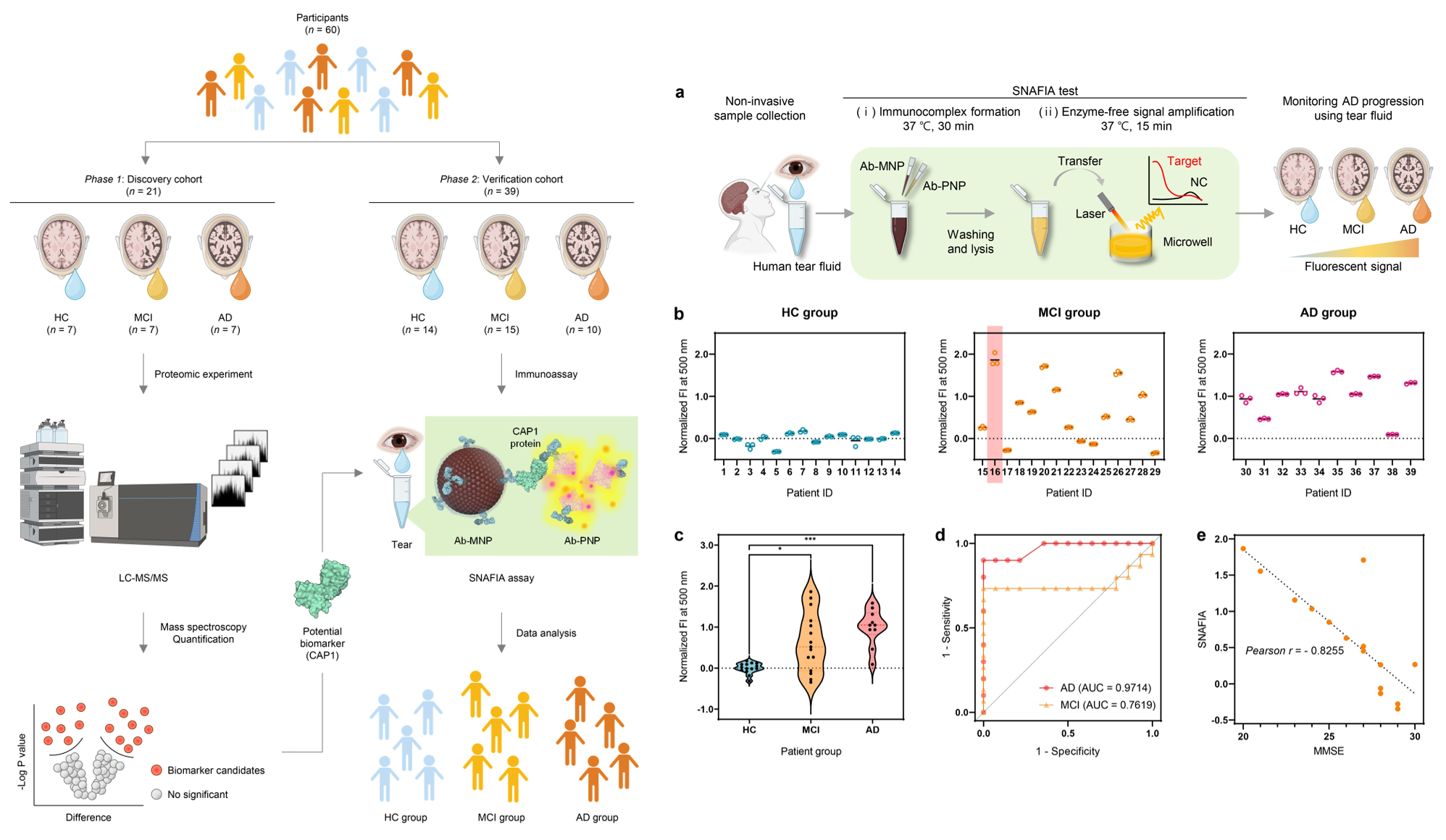
연세대 함승주 교수팀, 유/무기 나노 입자 기반의 고민감 진단 플랫폼 개발. 눈물 시료 내 표적 바이오마커 농축 기술과 신호 증폭 기술을 통합하여 알츠하이머 조기 진단 및 저비용 모니터링 가능성 확보 Professor Seungjoo Haam's team at Yonsei University developed an amplified fluorogenic immunoassay for early diagnosis and monitoring of Alzhei
연세대학교(총장 윤동섭) 화공생명공학과 함승주 교수 연구팀이 눈물 내 표적 단백질을 검출함으로서 알츠하이머 병 진단 뿐만 아니라 진행 단계를 모니터링 할 수 있는 유/무기 나노입자 기반의 면역분석법 (SNAFIA)을 개발했다. 알츠하이머병은 가장 흔한 신경 퇴행성 질환으로 뚜렷한 치료제가 없기 때문에 증상의 발병과 진행을 늦추는 효과를 극대화하는 조기 진단이 더욱 중요하다. 알츠하이머병 진단은 임상 징후와 증상의 관찰, 신경 인지 기능 검사, 초기 단계를 반영할 수 있는 알츠하이머병 촉진 바이오마커의 변화를 검사하는 것을 기반으로 한다. 뇌 기능 영상과 뇌척수액 분석 방법 등이 있으나 시간과 비용이 많이 들고 침습적 개입이 필요하며 부작용이 발생할 수 있기에 저비용, 저침습 진단법이 요구되고 있다. 연세대 함승주 교수팀이 개발한 센싱 플랫폼은 선정된 표적 단백질만을 선택적으로 검출할 수 있는 유/무기 나노 구조체 기반의 면역분석법으로, 자성 나노입자를 활용하여 표적 단백질을 분리 및 농축할 수 있는 시스템과 고분자 나노입자를 활용하여 일대다 염료 방출을 통한 신호 증폭 시스템의 통합으로 설계 되었다 . 연구팀은 고민감 신호 증폭 시스템 설계를 위해 친수성 내부와 소수성 막을 갖는 고분자 나노구조체를 합성하였으며 소수성 막 내부에 FRET 염료 분자를 담지하여 형광 표지제로 적용하였다. FRET 염료 분자들은 표적 단백질이 존재 하는 조건에서 방출되어 증폭된 형광 신호를 발생시키고 이외의 경우에는 소수성 막 내부에 갇혀 비특이 신호가 감소하도록 설계되었다. 실제로 상용화된 형광 표지제와 그 성능을 비교했을 때, 약 10 배 이상의 향상된 검출 수준을 보였으며 유/무기 나노입자 기반의 시스템 통합으로 높은 신호 대 잡음비 성능을 달성했다. 나아가 다양한 표적 단백질에 적용을 위해 맞춤형 항체로 표면 개질된 나노 구조체를 적용한 SNAFIA 테스트는, 실험실 플레이트 판독기를 사용하여 1 시간 이내에 아토몰 농도로 질병 관련 단백질을 검출할 수 있었으며, 액체 인간 생검에서 우수한 민감도와 선택도를 확보하였다. 특히, 이번 연구의 핵심은 39개의 임상 눈물 샘플을 대상으로 SNAFIA를 수행했을 때, 경도 인지 장애 집단과 알츠하이머 환자 집단이 건강한 집단에 비해 유의미한 신호 증가를 보인점에 있다. 아직 개념 증명 역할을 하는 수준이나, SNAFIA의 임상적 잠재력을 종합적으로 검증하기 위해서는 보다 광범위한 임상 조사가 필요하며 향후 대규모 임상 연구를 통해 견고한 결과를 얻어낸다면, SNAFIA는 간편하고 빠르며 눈물을 이용한 다양한 단백질 마커에 대해 높은 진단 정확도를 제공하므로 알츠하이머병 조기 진단을 위한 유망한 도구가 될 것으로 기대할 수 있다. 이번 연구는 보건복지부에서 주관한 보건의료기술연구개발 사업을 시작으로 과학기술정보통신부에서 주관하는 신변종감염병대응플랫폼핵심기술개발사업, 나노소재기술개발사업의 지원을 받아 함승주 교수 연구팀의 이소정 연구원(제 1저자), 용인 세브란스병원의 지용우 교수(공동 교신저자), 강남 세브란스병원의 조한나 교수(공동 교신저자)와 함께 진행됐으며, 세계적인 과학 분야 권위지 (IF=16.6) ‘Nature Communications’에 23년 12월 9일(현지시간) 게재됐다. A research team led by Prof. Seungjoo Haam of the Department of Chemical and Biological Engineering at Yonsei University (President Dong-seop Yoon) has developed an amplified fluorogenic immunoassay for early detection and monitoring of Alzheimer’s disease from tear fluid. Alzheimer's disease stands as the prevailing neurodegenerative disease with an absence of definitive remedies, accentuating the imperative for early detection to optimize therapeutic efficacy in mitigating symptom onset and progression. Diagnosis of Alzheimer's hinges upon meticulous clinical observation, neurocognitive assessment, and scrutiny of biomarkers implicated in its pathogenesis, offering insights into incipient stages. While conventional diagnostic modalities encompass brain imaging and cerebrospinal fluid analysis, their application is marred by protracted procedures, substantial costs, invasiveness, and potential adverse effects, underscoring the exigency for a cost-effective, minimally invasive diagnostic tools. The sensing platform developed by the team of Professor Seungjoo Haam at Yonsei University is an immunoassay based on organic and inorganic nanostructures that can selectively detect only selected target proteins, and is designed by integrating a system that can separate and concentrate target proteins using magnetic nanoparticles and a signal amplification system through one-to-many dye release using polymeric nanoparticles. To design the signal amplification system, the research team synthesized polymeric nanostructures with hydrophilic interior and hydrophobic membrane, and applied FRET dye molecules inside the hydrophobic membrane as fluorescent labels. The FRET dye molecules were designed to be released in the presence of the target protein, resulting in an amplified fluorescence signal, and to be confined inside the hydrophobic membrane in other cases, reducing the non-specific signal. In fact, when comparing its performance with commercially available fluorescent labeling agents, an improved detection level of about 10 times or more was obtained, and high signal-to-noise ratio performance was achieved by system integration based on organic and inorganic nanoparticles. Furthermore, the SNAFIA test using nanostructures surface-modified with antibodies for application to various target proteins was able to detect disease-related proteins at attomolar concentrations within 1 hour using a laboratory plate reader, and achieved excellent sensitivity and selectivity in liquid human biopsies. A key finding of the study was that when SNAFIA was performed on 39 clinical tear samples, the mild cognitive impairment group and the Alzheimer's patient group showed a significant increase in signal compared to the healthy group. While still serving as a proof of concept, more extensive clinical investigations are needed to comprehensively validate the clinical potential of SNAFIA, and if robust results are obtained in future large-scale clinical studies, SNAFIA could be a promising tool for early diagnosis of AD as it is simple, fast, and provides high diagnostic accuracy for a variety of protein markers using tears. This research was conducted by Dr. Sojeong Lee (first author) along with Prof. Seungjoo Haam’s research team, Prof. Yong Woo Ji (co-corresponding author), and Prof. Hanna Cho (co-corresponding author) with the support of the Korea Healthcare Technology Research and Development Project funded by the Ministry of Health and Welfare, the Nanomaterial Technology Development Project and Emerging Infectious Disease Response Platform Core Technology Development Project promoted by the Ministry of Science and ICT. The work was published on December 9, 2023 (local time) in the prestigious journal of ‘Nature communications’. Nat. Commun. (2023) (IF: 16.6)Published: December 09, 2023https://doi.org/10.1038/s41467-023-43995-5
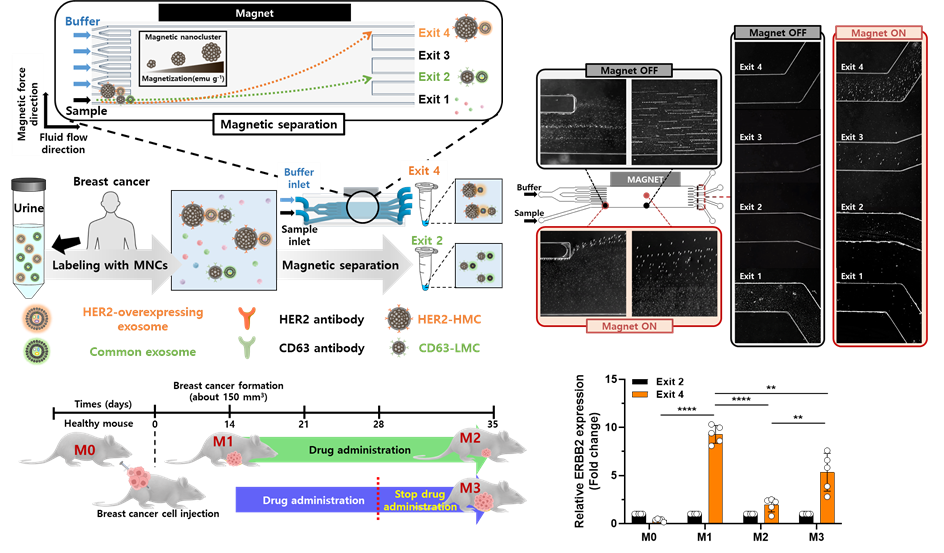
연세대 함승주 교수팀, 자성 입자 활용, 자화도 차이에 따른 유방암 특이적 엑소좀 (HER 2 positive exosome) 다중 분리 칩 기술 개발. Professor Seungjoo Haam's team at Yonsei University developed a microfluidic chip for HER2-Positive cancer-derived exosomes isolation and detection
연세대학교(총장 서승환) 화공생명공학과 함승주 교수 연구팀이 자성 입자 활용하여 다중 분리를 통해 HER2 과발현 유방암 유래 엑소좀을 분리 및 검출할 수 있는 미세 유체 칩을 개발했다. 엑소좀은 세포 내에서 생성 되어 외부로 방출되는 소포밖 소포체 중 하나이다. 엑소좀은 30-150 나노미터 (nm) 크기로 모세포의 단백질, RNA, DNA 등의 다양한 생체물질을 포함하고 있으며 매우 안정적으로 유지된다. 체액 속의 엑소좀 분석은 최근 혁신적이고 유망한 액체 생검 방법으로 최근 주목 받고 있으나, 기존 방식으로는 분석에 적합한 농도의 엑소좀을 체액 속의 다양한 불순물로부터 얻는 것이 어렵다. 연세대 함승주 교수팀이 개발한 미세 유체 칩은 자성 나노입자를 이용하여 다종 엑소좀을 서로 다른 자화도로 표지하고, 미세유체 칩 내부에서 자화도 차이에 따라 다중 분리한다. 이를 통해 체액 속에서 질환 유래 엑소좀과 비표적 엑소좀을 분리할 수 있어, 질환 유래 엑소좀을 통해 정확한 질환 진단이 가능하며 비표적 엑소좀을 내부통제군(internal control)으로 활용하여 효과적인 모니터링이 가능하다. 연구팀은 다양한 자화도를 나타내는 자성 나노입자를 합성하고, 이에 질환 유래 엑소좀과 비표적 엑소좀에 표지되도록 항체를 결합하였다. 미세유체 칩 내부에서 외부 자기장에 의한 자화 표지된 엑소좀의 거동을 최적화하고 분리 효율에 대해 확인하였다. 소변 내 HER2 과발현 엑소좀과 비표적 엑소좀을 동시에 분리하여 치료 효과에 대한 실시간 모니터링에 대해 입증하였다. 이 연구를 통해 체액으로부터 분리한 엑소좀으로 암 진단 및 약물 치료 효과 모니터링이 가능할 것으로 기대되며, 바이오마커 선정에 따라 암 뿐만 아니라 다양한 난치성 질환에 활용 가능할 것으로 예상된다. 이번 연구는 과학기술정보통신부에서 주관하는 나노소재기술개발사업, 바이오·의료기술개발사업, 신변종감염병대응플랫폼핵심기술개발사업, 환경부에서 추진하는 생물학적위해인자관리기술개발사업의 지원으로 함승주 교수 연구팀의 문병걸 연구원(제1저자), 한국생명공학연구원의 임은경 박사(공동 교신저자)과 함께 진행됐으며, 세계적인 과학 분야 권위지 ‘바이오 센서 앤 바이오 일렉트로닉스 (Biosensors & Bioelectronics)’에 11월 1일자(현지시간)로 게재됐다. Yonsei University’s (President Seung-Hwan Seo) department of chemical and biomolecular engineering Professor Seungjoo Haam's research team developed an immuno-magnetophoresis-based microfluidic chip to isolate and detect HER2-Positive cancer-derived exosomes via multiple separation. Exosomes are extracellular vesicles produced inside cells and released to the outside.Exosomes are a type of endoplasmic reticulum that is produced inside the cell and released to the outside. Exosomes are 30-150 nanometers (nm) in size and contain various biological substances from the parent cells, such as proteins, RNA and DNA, and are maintained in a very stable form. The analysis of exosomes in body fluids has recently attracted attention as an innovative and promising liquid biopsy method. However, obtaining exosomes of appropriate concentration for analysis from contaminants has been a limitation of existing methods. The microfluidic chip developed by Professor Ham Seung-joo's team at Yonsei University uses magnetic nanoparticles to label multiple types of exosomes with different degrees of magnetization, and separates them multiple times according to the differences in magnetization within the microfluidic chip. This allows disease-derived exosomes and non-target exosomes to be separated in body fluids, enabling accurate disease diagnosis by disease-derived exosomes and effective monitoring by using non-target exosomes as an internal control group. The research team synthesized magnetic nanoparticles with different degrees of magnetization and combined them with antibodies to label disease-derived exosomes and non-target exosomes. The behavior of the magnetically labeled exosomes was optimized by an external magnetic field inside the microfluidic chip and the separation efficiency was confirmed. Real-time monitoring of treatment effect was demonstrated by simultaneous separation of HER2-overexpressing exosomes and non-targeted exosomes in urine. This study is expected to enable cancer diagnosis and monitoring of drug treatment effects using exosomes isolated from body fluids, and depending on the selection of biomarkers, it is expected to be used not only for cancer but also for various incurable diseases. This research was conducted with Researcher Byeonggeol Mun (first author) of Professor Seungjoo Haam's research team, and Dr. Eun-Kyung Lim (co-corresponding author) with the support by the Ministry of Science and ICT and Ministry of Environment. The work was published in the prestigious journal of 'Biosensors & Bioelectronics’ on November 1 (local time). Biosensors and Bioelectronics (2023) (IF: 12.625)Published: november 01, 2023https://doi.org/10.1016/j.bios.2023.115592
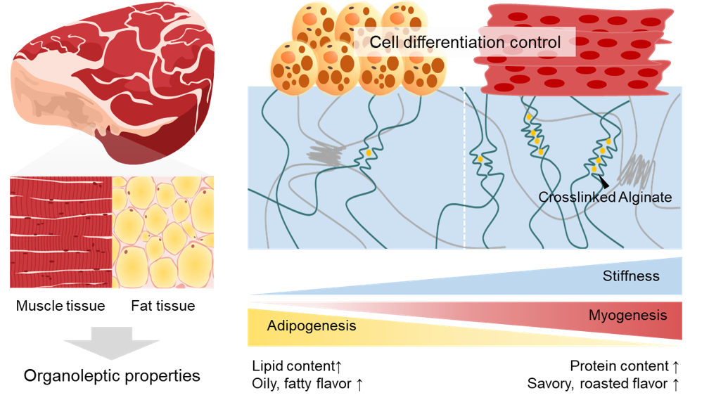
연세대 홍진기 교수팀, 알긴산의 가교 조절을 통해 등심과 안심의 관능 특성을 구현한 배양육 소재 개발 Professor Jinkee Hong's team at Yonsei University developed cultured meat materials embodying the organoleptic properties of sirloin and tenderloin through controlling the cross-linking of alginic
연세대학교(총장 서승환) 화공생명공학과 홍진기 교수 연구팀이 천연 고분자의 가교 조절을 통해 다양한 도축 부위의 관능 특성을 구현할 수 있는 배양육 소재를 개발했다. 배양육은 실험실에서 인공적으로 생산하는 육류로, 도축과 자원 소모를 최소화할 수 있는 지속 가능한 육류로 떠오르고 있다. 배양육의 궁극적인 목표는 도축육이 갖는 다양한 물리적, 생물학적 특성을 모사하여 체외 배양으로 도축육의 감각적 특성을 달성하는 것이다. 그러나 아직까지 세포 배양을 통해 여러 조직이 결합되어 복합적인 향미와 식감을 발현하는 도축육의 관능적인 특징을 구현하는 데에는 한계가 있다. 이에 연세대 홍진기 교수 연구팀은 콜라겐 유래 고분자인 젤라틴과 갈조류 유래 다당류인 알긴산으로 구성된 세포 배양 지지체를 개발했다. 특히 알긴산의 카르복실기와 양이온이 만나 이온결합을 형성하여 알긴산 사슬의 구조 변화가 나타나는 .점을 활용해 지지체의 기계적 강도를 달리하였다. 알긴산의 이온 가교도가 조절되면 하이드로겔의 기계적 강도가 달라지는데, 이를 이용하여 근육 조직의 영률 (~12 kPa)에 도달한 지지체와 지방 조직의 영률 (~3 kPa)에 도달한 지지체를 제조하고, 근육 세포와 지방 세포의 분화 거동을 조절하였다. 도축육의 식감과 향미 등의 관능적 특성과 영양항적 특성은 근조직과 지방 조직의 생물학적 특성에서 기인하기 때문에, 연세대 홍진기 교수 연구팀은 지지체를 이용해 조절된 세포의 분화 거동이 다양한 관능 및 영양학적 특성에도 영향을 줄 것으로 가정하였다. 결과적으로, 물성에 따라 근분화와 지방분화 정도가 다른 배양육을 제조하였을 때 식감과 향미, 영양학적 특성이 유의미하게 달라지며 두 세포의 분화 정도가 최대화된 배양육에서 도축 소고기의 특성이 나타남을 확인하였다. 이러한 결과를 바탕으로, 근분화가 증가된 배양육과 지방분화가 증가된 배양육의 공유 결합을 유도하여 다양한 도축 부위의 관능 특성을 구현할 수 있는 배양육 제조 기술을 개발했다. 연세대 홍진기 교수 연구팀은 이번 연구가 기존 배양육 연구에서 집중하지 않았던 도축육의 관능 특성 구현을 최초로 보고하였다는 점에서 차별성이 있으며, 향후 배양육 생산에 폭넓게 적용될 수 있는 초석기술로 기대한다고 밝혔다. 연세대 홍진기 교수는 “이번 연구는 고분자 기반 소재의 특성을 조절하는 화학공학 기술이 생명공학 기술, 식품 공학 기술과 융합되어 미래 식품 분야에도 적용될 수 있음을 보여주었다”고 전하며, “앞으로도 우리 연구팀의 다양한 소재 기술을 활용해 배양육 산업의 발전에 기여할 수 있는 연구를 이어 나갈 것" 이라고 덧붙였다. 본 연구는 대한민국 산업통상자원부(MOTIE), 한국연구재단(NRF), 국방기술진흥연구소의 지원을 받아 수행되었다. 이번 연구는 홍진기 교수 연구팀의 이미래 박사과정생(제 1저자)가 진행했으며, 국제 학술 권위지 ‘네이처 커뮤니케이션즈(Nature Communications)’에 1월 2일자(현지시간)로 게재됐다.A research team led by Prof. Jinkee Hong of the Department of Chemical and Biomolecular Engineering at Yonsei University (President Seung-Hwan Seo) has developed a cultured meat material that can mimic the sensorial characteristics of various slaughtered cuts of meat through controlling the cross-linking degree of alginic acid. Cultured meat, produced artificially in laboratories, is emerging as a sustainable meat that minimizes slaughter and resource consumption. The ultimate goal of cultured meat is to achieve the sensorial characteristics and nutritional value of slaughtered meat through in vitro cultivation, mimicking the diverse physical and biological properties of slaughtered meat. However, there are limitations in replicating these features of slaughtered meat through cell cultivation, because the properties of slaughtered meat involve the complex interplay of various tissues. To address this challenge, Prof. Jinkee Hong’s research team at Yonsei University developed a cell culture scaffold composed of collagen-derived polymer gelatin and alginic acid, a polysaccharide derived from algae. They specifically manipulated the mechanical strength of the scaffold by utilizing the interaction between carboxyl groups in alginic acid and cations, causing structural changes in the alginic acid chain. By adjusting the ionic cross-linking of alginic acid, they developed the scaffolds reaching the mechanical stiffnesses of muscle tissue (~12 kPa) and fat tissue (~3 kPa). Using these scaffolds, the differentiation behaviors of bovine muscle cells and fat cells were precisely regulated. Since the organoleptic and nutritional characteristics of slaughtered meat originate from the biological properties of muscle and fat tissues, the research team hypothesized that the controlled differentiation behavior of cells using the scaffolds would impact various sensorial and nutritional attributes. Consequently, they confirmed significant differences in texture, flavor, and nutritional characteristics when producing cultured meat with different degrees of muscle and fat differentiation. Finally, covalent cross-linking was induced between the cultured meats containing increased muscle differentiation and increased fat differentiation, resulting a novel assembled cultured meat which can mimic the organoleptic characteristics of diverse slaughtered meat cuts. Prof. Jinkee Hong stated, "This study demonstrates the integration of chemical engineering technology in adjusting the properties of polymer-based materials with the fields of life sciences and food engineering. It highlights the potential application of our diverse material technologies to contribute to the advancement of the cultured meat industry.“ This research was conducted with the support of the Ministry of Trade, Industry, and Energy (MOTIE), the National Research Foundation (NRF), and the Agency for Defense Development. The study was conducted by Milae Lee (first author, PI: Prof. Jinkee Hong) and was published in the prestigious journal, Nature Communications, on January 2nd, 2024 (local time).Nature Communications (2023) (IF: 16.6)Published: January 2nd, 2024https://www.nature.com/articles/s41467-023-44359-9
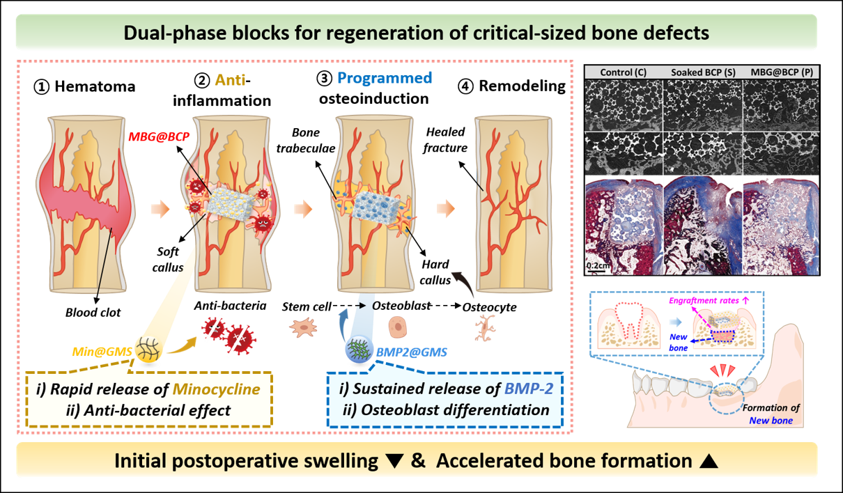
연세대 홍진기 교수팀, 임계 크기의 골 결손 치료를 위한 다중 약물 방출 블록 기반 플랫폼 개발. Professor Jinkee Hong’s group at Yonsei University developed a novel multi-drug release dual-phase blocks for regeneration of critical-sized bone defects.
연세대학교(총장 윤동섭) 화공생명공학과 홍진기 교수 연구팀이 임계 크기의 골 결손 (critical-sized bone defect)의 효과적인 치료를 위하여 다중 약물을 방출하는 블록 기반 플랫폼을 개발하였다. 임계 크기의 골 결손이 발생하면, 임플란트와 골 대체제를 이용하여 치조골 재생 능력을 높이는 치료가 필요하다. 특히, 골재생 분야에서 제2형 골현성단백질 (bone morphogenetic protein-2, BMP-2)라고 불리는 성장인자를 도입하여 임상적으로 골이식술, 골유도재생술 등 여러 술식에서 이용하고 있다. 그러나, 기존 BMP-2의 전달 방식은 대부분 이식 후 과잉 방출되는 등 제어되지 않고 불규칙하여 심한 부종, 신생물, 낭종 형성과 같은 조직 형성 및 파골세포 흡수를 포함한 임상적 합병증을 유발할 수 있어 임상적 사용이 제한적이다. 또한, 이러한 성장인자의 방출이 이식 직후인 염증 단계 (inflammatory phase)에 대부분 이루어지면서 성장인자의 효과를 감소시킨다. 연세대 홍진기 교수팀이 이와 같은 문제를 해결하고자 개발한 블록 기반 플랫폼은 이상 인산칼슘 (biphasic calcium phosphate, BCP) 블록 내부에 약물이 담지되어 있는 젤라틴 마이크로 입자를 탑재하고, 블록 표면을 젤라틴 기반 필름으로 코팅하여 약물 방출을 조절함으로써 효과적인 골 형성 효과를 보여주었다. 연구팀은 젤라틴 마이크로 입자의 분해도를 조절하여 최적의 항생제와 성장 인자 방출 거동을 보이는 입자를 설계하였다. 블록 이식 초반에는 염증 반응을 줄이기 위하여 분해 속도가 빠른 젤라틴 마이크로 입자에서 항생제가 신속하게 방출되고, 이식 후에는 분해 속도가 느린 젤라틴 마이크로 입자에서 성장 인자가 지속적으로 방출되어 성공적인 골 재생 효과를 입증하였다. 최종적으로, 해당 플랫폼의 성능을 하악 결손 동물실험 모델에서 검증함으로써, 항생제와 성장인자의 치료 단계에 따른 순차적인 방출을 통해 성공적인 골 재생 효과를 확인하였다. 특히, 초기 항생제로 인한 염증 반응의 감소로 인해 수술 후 초기 부종이 줄어들었고, 이후 지속적인 성장인자의 방출로 인해 신생골 형성을 촉진하여 기존 치료법으로는 치료할 수 없었던 큰 부피의 골 결손도 치료할 수 있는 가능성을 제시하였다. 연세대 홍진기 교수는 “해당 연구를 통해 항생제와 성장인자의 순차적 치료가 골 재생에 미치는 효과를 입증함으로써, 하악 결손 동물 모델에서의 새로운 치료 전략을 제시하였다. 이러한 결과는 골 결손 치료에 대한 새로운 지평을 열면서 동시에 미래의 임상 응용에 대한 중요한 기초를 제공할 것으로 기대된다“ 고 전했다. 본 연구는 한국연구재단 (NRF), 국가신약개발재단 (KDDF), 한국보건산업진흥원 (KHIDI)의 지원을 받아 수행되었다. 이번 연구는 홍진기 교수 연구팀의 김지유 박사과정생(제1저자), 박소현 박사(제1저자)가 연세대학교 치과대학 박진영 교수(제1저자)와 함께 진행하였으며, 세계적인 과학 분야 권위지 ‘나노 투데이 (Nano Today)’에 1월 5일자(현지시간)로 온라인 게재됐다.Yonsei University’s (President Dong-Sup Yoon) department of chemical and biomolecular engineering Professor Jinkee Hong's research team developed a novel multi-drug release dual-phase blocks for regeneration of critical-sized bone defects. When a critical size bone defect occurs, treatment to increase the alveolar bone regeneration ability using implants and bone replacements is required. In particular, a growth factor called bone morphogenetic protein-2 (BMP-2) has been introduced in the field of bone regeneration and is clinically used in various procedures such as bone transplantation and bone-induced regeneration. However, most of the existing delivery methods of BMP-2 are uncontrolled and irregular, such as excessive release after transplantation, which can lead to clinical complications including severe edema, neoplasia, tissue formation such as cyst formation, and osteoclast uptake, so its clinical use is limited. In addition, the release of these growth factors mostly takes place in the inflammatory phase immediately after transplantation, reducing the effect of growth factors. The dual-phase block platform developed by Professor Jinkee Hong’s team at Yonsei University to solve this problem has shown an effective bone formation effect by loading gelatin microparticles containing drugs inside a block of biophilic calcium phosphate (BCP), and coating the surface of the block with a gelatin-based film to control drug release. The research team designed particles showing optimal antibiotic and growth factor release behavior by controlling the decomposition of gelatin microparticles. In order to reduce the inflammatory response at the beginning of block transplantation, antibiotics were quickly released from gelatin microparticles with a high decomposition rate, and after transplantation, growth factors were continuously released from gelatin microparticles with a slow decomposition rate, demonstrating a successful bone regeneration effect. Finally, the performance of the platform was verified in the mandibular defect animal experimental model, and the successful bone regeneration effect was confirmed through the sequential release of antibiotics and growth factors according to the treatment stage. In particular, the initial swelling decreased after surgery due to the decrease in the inflammatory response due to the initial antibiotic, and the possibility of treating even large-volume bone defects that could not be treated with conventional treatments was suggested by promoting nephrotic bone formation due to the continuous release of growth factors. Professor Jinkee Hong at Yonsei University said, "By demonstrating the effect of sequential treatment of antibiotics and growth factors on bone regeneration through this study, we presented a new treatment strategy in the mandibular defect animal model. These results are expected to open new horizons for the treatment of bone defects while providing an important basis for future clinical applications.“ This research was conducted with the support of the National Research Foundation (NRF), Korea Drug Development Fund (KDDF), and Korea Health Industry Development Institute (KHIDI). The study was conducted by Jiyu Kim (first author) and Dr. Sohyeon Park (first author) with Prof. Jin-Young Park (first author) of Yonsei University College of Dentistry, and published online in the prestigious scientific journal ‘Nano Today' on January 5th (local time). Nano Today (2024) (IF: 17.4)Published: January 05, 2024https://doi.org/10.1016/j.nantod.2023.102120
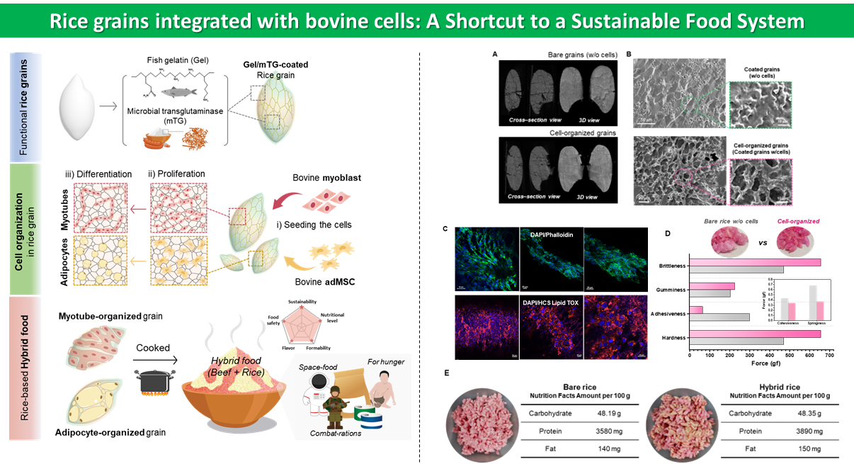
연세대 홍진기 교수팀, 지속 가능한 식량 시스템을 구축하기 위한 쌀알 기반 배양육 개발 - 나노 코팅과 쌀알, 그리고 가축 세포의 통합이 새로운 하이브리드 식품의 탄생을 이끌어 - - 국제적 최고 권위 저널 ‘Matter (IF 18.9)’게재 -
사진 1. (왼쪽) 연세대학교 화공생명공학과 홍진기 교수, (오른쪽) 연세대학교 화공생명공학과 박소현 박사 (1저자) 사진 2. 코팅된 쌀알과 가축 소 세포를 통합하여 쌀알 기반의 배양육을 제조하는 전략을 보여주는 개략도 및 개발된 쌀알 배양육의 특징을 보여주는 대표 결과 연세대학교 홍진기 교수(화공생명공학과) 연구팀은 지속 가능한 식품 시스템을 위해 나노 코팅으로 기능화된 쌀알에 가축 세포를 통합하여 영양이 풍부한 쌀알 기반 배양육을 개발하였다. 또한, 이 새로운 배양육의 식품 특성과 생산 가치에 대해 논의하여 식량 위기와 지구 온난화로부터 지속 가능한 식품으로서의 잠재력을 보고하였다. 건강 문제 증가, 전염병 위험, 기후 변화, 자원 부족 등의 요인으로 인해 식품 시스템은 전 세계적으로 구조적인 변화를 겪고 있다. 따라서 피할 수 없는 식량 위기에 대비하여 안정적인 식량 체계를 확보하는 것은 오늘날 인류의 숙원사업이다. 이에 따라 인공육, 곤충 유래 단백질, 및 3D 프린팅 식품 등의 흥미로운 미래 식품이 최근까지도 계속 보고되고 있다. 그러나 지속 가능한 식품 시스템을 이루기 위해서는 제품 생산과정의 안전성과 안정성이 보장되어야 하며, 제조된 식품은 영양이 균일하고 높은 가공성을 가져야 한다. 안타깝게도 지금까지 보고된 미래 식품 후보들은 영양 불균형, 생소한 맛, 열악한 성형 및 가격 경쟁력 등 상품화 측면에서 실질적인 한계를 갖고 있다. 연구진은 일상에서 쉽게 접할 수 있으며 필수 영양소를 함유한 쌀알을 가축 세포의 3D 지지체로 사용하여 상용화 가능성이 높은 새로운 형태의 하이브리드 식품을 제조하는 전략을 설계했다. 쌀알의 패킹 구조는 넓은 표면적과 다공성 및 조직화 된 공간을 제공하여 가축 세포의 함입을 수용하며, 쌀알을 생선 젤라틴과 식품 등급 효소로 구성된 나노 코팅으로 기능화함으로써 쌀알의 세포 수용량을 크게 증가시킬 수 있다. 가축 소의 근아세포와 지방유래 중간엽 줄기세포를 코팅된 곡물 위에 증식 및 분화시켜 조직화 된 세포를 함유한 영양이 풍부한 쌀알 배양육이 탄생되었다. 쌀알 배양육은 일반 쌀알과 비교하여 뚜렷한 형태적, 기계적 특성을 나타냈으며, 이러한 차이는 밥의 식감에 큰 영향을 미쳤다. 쌀알 배양육의 영양성분, 식감 및 향미 분석 등의 결과는 이의 영양학적 가치와 식품 잠재력을 증명했다. 쌀알 배양육은 일반 쌀보다 더 많은 단백질과 지방을 함유하고 있어 풍부한 풍미를 나타냈다. 또한, 쌀알 배양육의 단백질은 소 조직 단백질과 유전적으로 18.54% 일치하였다. 홍진기 교수는 이 전략이 식품, 지지체 및 세포가 상호 이익이 되는 하이브리드 기술이며, 재료 간의 상호작용을 최적화하여 다른 식품 성분에도 폭넓게 적용할 수 있다고 밝혔다. 더불어 미래 식품 개발에 재료 공학을 도입하면 다양한 형태의 미래 식품 개발이 실현 가능할 것이라 기대했다. 또한, 본 기술은 자가 생산이 가능한 식량 체계에 적용 가능하므로 이러한 곡물 기반 단백질원은 저개발국, 전쟁 및 우주 등 비상사태에 대응한 구호 식량으로 개발될 수 있다고 평가했다. 본 연구는 한국연구재단(NRF)와 한국보건산업진흥원(KHIDI) 사업의 지원을 받아 진행되었다. 연구 결과는 재료 분야 국제 최고 권위 학술지 “매터 (Matter)에 2024년 2월 14일 게재되었다. 논문 제목: Rice grains integrated with animal cells: A shortcut to a sustainable food system
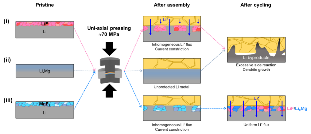
연세대 정윤석 교수팀, 전고체전지 리튬금속 보호층 적용 메커니즘 제시 무기물, 금속, 복합 보호층 적용 리튬금속 음극 거동 분석 장수명 리튬금속 전고체전지 구현 난제 해경방안 제시. Professor Yoon Seok Jung's team at Yonsei University presented a new mechanism regarding the application of interlayer for Lithium metal all-solid-state
연세대학교(총장 서승환) 화공생명공학과 정윤석 교수 연구팀은 전고체전지 리튬금속 음극의 무기물, 금속, 그리고 복합 보호층을 적용한 전고체전지 제작 및 구동시 거동을 분석하고, 이를 이용하여 고안정성, 장수명 리튬금속 전고체전지 기술을 개발했다. 현재 상용화된 리튬이온전지는 인화성의 유기계 액체전해질을 사용하기 때문에 발화 및 폭발에 대해 매우 취약하여 안전성 및 안정성에 한계가 있다. 또한, 이론 용량이 적은 흑연 음극을 사용하기에 에너지밀도 향상에도 어려움이 있다. 이에, ‘꿈의 배터리’로 불리는 전고체전지가 큰 주목을 받고 있다. 전고체전지는 무기물 기반의 고체전해질을 사용함으로써 안전성/안정성을 확보하고, 이론 용량이 높은 리튬금속 음극을 적용함으로써 에너지밀도를 획기적으로 개선할 수 있다. 황화물계 고체전해질은 상온에서 높은 이온전도 특성(1~10mS/cm)을 가지며 무른 기계적 물성으로 고체-고체간 접촉면 형성이 용이해 핵심 무기계 고체전해질소재로 개발되고 있다. 하지만, 리튬금속 적용 시 접촉 계면에서 극심한 부반응을 일으키고, 리튬 수지상 성장으로 인한 내부단락 발생 등의 문제가 발생한다. 지금까지, 해결방안으로 무기물, 금속, 또는 둘을 합친 복합 보호층 적용에 관한 연구들이 많이 진행되었다. 하지만, 강한 압력이 가해지는 전고체전지 조립 과정부터 부피변화를 수반하는 충/방전 과정에 이르는 동안에 보호층의 물리적, 전기화학적 거동에 대한 이해는 거의 전무한 상황이었다. 연세대 정윤석 교수 연구팀은 대표적인 보호층 소재인 무기물(LiF), 금속(Mg), 복합 보호층(MgF2)이 적용된 리튬금속 음극의 거동을, 보호층 적용부터 전고체전지 조립, 그리고 충방전에 이르는 전 과정에 걸쳐 종합적으로 분석했다. 그 결과, 무기물 코팅층은 전고체전지 조립시 가해지는 외부 압력에 의해 물리적으로 붕괴됐고, 금속 보호층의 경우 리튬금속 내부로 확산하여 보호층으로 기능을 하지 못하는 한계점을 발견했다. 하지만 복합 보호층의 경우에는 초기 충방전 과정에서 일어나는 화학적 변환반응을 (xLi + MgF2 → LixMg + LiF) 통해 보호층 형상을 회복하여 안정적인 리튬금속 전탈착 거동을 보였다. 정윤석 교수는 “이번 연구는 그동안 전고체전지 리튬금속 보호층 연구에서 간과되었던 전고체전지 제작시에 발생할 수 있는 물리적 결함에 대해 지적하며 보호층 연구에 있어 새로운 차원을 제시한 결과로, 전고체전지 상용화에 기여할 수 있을 것이라 기대한다”고 전했다. 본 연구는 산업통상자원부 및 산업기술평가관리원(KEIT) 그리고 연구재단(NRF)의 연구비 지원으로 정윤석 교수(교신저자)와 임해찬나라 박사과정생(제1저자) 등이 함께 연구를 수행했고, 에너지기술 분야 국제 저명 학술지 ‘어드벤스드 에너지 머티리얼즈(Advanced Energy Materials)’에 게재됐다(논문명: Rationally Designed Conversion-type Lithium Metal Protective Layer for All-Solid-State Lithium Metal Batteries). The currently commercialized lithium-ion batteries, due to their use of flammable organic liquid electrolytes, are highly susceptible to ignition and explosion, posing limitations on safety and stability. Additionally, the use of graphite anodes with low theoretical capacity makes it challenging to improve energy density. In response to these challenges, there is growing attention on the all-solid-state batteries, known as "Dream battery". Solid-state batteries utilize inorganic solid electrolytes to ensure safety and stability, and they employ lithium metal anodes with high theoretical capacity to significantly enhance energy density. Sulfide-based solid electrolytes exhibit high ion conductivity (1~10 mS/cm) at room temperature and have ductile mechanical properties, making them promising materials for solid-solid contact interfaces. However, issues arise when applying lithium metal, such as severe reactions at the contact interface and internal short circuits due to lithium dendrite growth. Various studies have been conducted to address these challenges, focusing on the application of inorganic, metal, or composite protective layers. However, understanding the physical and electrochemical behaviors of protective layers during the entire process—from the assembly of solid-state batteries under high pressure to the volume changes during charge/discharge cycles—has been largely lacking. Professor Jung Yoon-seok's research team at Yonsei University comprehensively analyzed the behavior of lithium metal anodes coated with representative protective layers, including inorganic (LiF), metal (Mg), and composite protective layers (MgF2). The results revealed that the inorganic coating layer collapsed physically under external pressure during the assembly of solid-state batteries, while the metal protective layer diffused into the lithium metal, limiting its protective function. In contrast, the composite protective layer exhibited stable behavior, recovering its protective layer shape through a chemical transformation reaction (xLi + MgF2 → LixMg + LiF) during the initial charge/discharge cycles. Professor Jung Yoon-seok emphasized that this research sheds light on the often-overlooked physical defects that can occur during the fabrication of solid-state batteries and introduces a new dimension to protective layer research. He expressed hope that this study could contribute to the commercialization of solid-state batteries. This research was conducted by Haechannara Lim (first author) of Professor Yoon Seok Jung’s research team at Yonsei University with the support of Ministry of Science and ICT’s original technology development project (The program of phased development of carbon neutral technologies), and technology innovation program funded by the Ministry of Trade, Industry and Energy (MOTIE, Korea) and Korea Planning & Evaluation Institute of Industrial Technology (KEIT). The research has been recently accepted to Advanced Energy Materials, an internationally renowned academic journal in the field of energy. (Article title : Rationally Designed Conversion-type Lithium Metal Protective Layer for All-Solid-State Lithium Metal Batteries). Advanced Energy Materials (2024) (IF: 27.8)Published: 01. 18, 2024https://doi.org/10.1002/aenm.202303762
1
2
3
4